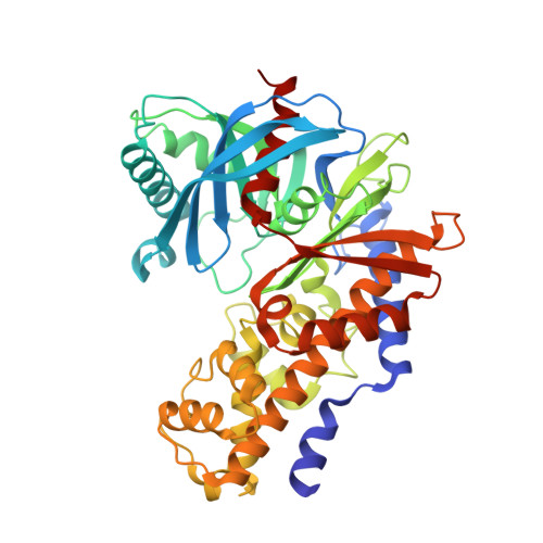
3A0I

Human glucokinase in complex with a synthetic activator


Experimental Data Snapshot

  • Method: X-RAY DIFFRACTION
  • Resolution: 2.20 Å
  • R-Value Free: 0.277 
  • R-Value Work: 0.226 
  • R-Value Observed: 0.229 

wwPDB Validation   3D Report Full Report


Ligand Structure Quality Assessment 


This is version 1.4 of the entry. See complete history


Literature

Discovery of novel 3,6-disubstituted 2-pyridinecarboxamide derivatives as GK activators

Mitsuya, M.Kamata, K.Bamba, M.Watanabe, H.Sasaki, Y.Sasaki, K.Ohyama, S.Hosaka, H.Nagata, Y.Eiki, J.Nishimura, T.

(2009) Bioorg Med Chem Lett 19: 2718-2721

  • DOI: https://doi.org/10.1016/j.bmcl.2009.03.137
  • Primary Citation of Related Structures:  
    3A0I, 3GOI

  • PubMed Abstract: 

    A novel class of 3,6-disubstituted 2-pyridinecarboxamide derivatives was designed based on X-ray analysis of the 2-aminobenzamide lead class. Subsequent chemical modification led to the discovery of potent GK activators which eliminate potential toxicity concerns associated with an aniline group of the lead structure. Compound 7 demonstrated glucose lowering effect in a rat OGTT model.


  • Organizational Affiliation

    Banyu Tsukuba Research Institute, Banyu Pharmaceutical Co. Ltd, Okubo, Tsukuba, Ibaraki, Japan. morihiro_mitsuya@merck.com


Macromolecules
Find similar proteins by:  (by identity cutoff)  |  3D Structure
Entity ID: 1
MoleculeChains Sequence LengthOrganismDetailsImage
GlucokinaseA [auth X]455Homo sapiensMutation(s): 0 
EC: 2.7.1.2
UniProt & NIH Common Fund Data Resources
Find proteins for P35557 (Homo sapiens)
Explore P35557 
Go to UniProtKB:  P35557
PHAROS:  P35557
GTEx:  ENSG00000106633 
Entity Groups  
Sequence Clusters30% Identity50% Identity70% Identity90% Identity95% Identity100% Identity
UniProt GroupP35557
Sequence Annotations
Expand
  • Reference Sequence
Small Molecules
Ligands 3 Unique
IDChains Name / Formula / InChI Key2D Diagram3D Interactions
AJI
Query on AJI

Download Ideal Coordinates CCD File 
C [auth X]3-[(4-fluorophenyl)sulfanyl]-N-(4-methyl-1,3-thiazol-2-yl)-6-[(4-methyl-4H-1,2,4-triazol-3-yl)sulfanyl]pyridine-2-carboxamide
C19 H15 F N6 O S3
SJTQKYKXCYVFHX-UHFFFAOYSA-N
GLC
Query on GLC

Download Ideal Coordinates CCD File 
B [auth X]alpha-D-glucopyranose
C6 H12 O6
WQZGKKKJIJFFOK-DVKNGEFBSA-N
NA
Query on NA

Download Ideal Coordinates CCD File 
D [auth X]SODIUM ION
Na
FKNQFGJONOIPTF-UHFFFAOYSA-N
Binding Affinity Annotations 
IDSourceBinding Affinity
AJI BindingDB:  3A0I EC50: 250 (nM) from 1 assay(s)
Experimental Data & Validation

Experimental Data

  • Method: X-RAY DIFFRACTION
  • Resolution: 2.20 Å
  • R-Value Free: 0.277 
  • R-Value Work: 0.226 
  • R-Value Observed: 0.229 
  • Space Group: P 65 2 2
Unit Cell:
Length ( Å )Angle ( ˚ )
a = 79.732α = 90
b = 79.732β = 90
c = 321.975γ = 120
Software Package:
Software NamePurpose
REFMACrefinement
WEISdata scaling
HKL-2000data reduction
HKL-2000data scaling

Structure Validation

View Full Validation Report


Ligand Structure Quality Assessment 


Entry History 

Deposition Data

Revision History  (Full details and data files)

  • Version 1.0: 2009-04-28
    Type: Initial release
  • Version 1.1: 2011-07-13
    Changes: Version format compliance
  • Version 1.2: 2017-10-11
    Changes: Refinement description
  • Version 1.3: 2020-07-29
    Type: Remediation
    Reason: Carbohydrate remediation
    Changes: Data collection, Derived calculations, Structure summary
  • Version 1.4: 2023-11-01
    Changes: Data collection, Database references, Refinement description, Structure summary