2Y6I

Crystal Structure of Collagenase G from Clostridium histolyticum in complex with Isoamylphosphonyl-Gly-Pro-Ala at 3.25 Angstrom Resolution


Experimental Data Snapshot

  • Method: X-RAY DIFFRACTION
  • Resolution: 3.25 Å
  • R-Value Free: 0.266 
  • R-Value Work: 0.216 
  • R-Value Observed: 0.219 

wwPDB Validation   3D Report Full Report


This is version 1.3 of the entry. See complete history


Literature

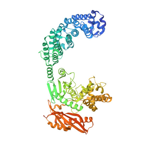
Structure of Collagenase G Reveals a Chew-and -Digest Mechanism of Bacterial Collagenolysis

Eckhard, U.Schoenauer, E.Nuess, D.Brandstetter, H.

(2011) Nat Struct Mol Biol 18: 1109

  • DOI: https://doi.org/10.1038/nsmb.2127
  • Primary Citation of Related Structures:  
    2Y3U, 2Y50, 2Y6I, 2Y72

  • PubMed Abstract: 

    Collagen constitutes one-third of body protein in humans, reflecting its extensive role in health and disease. Of similar importance, therefore, are the idiosyncratic proteases that have evolved for collagen remodeling. The most efficient collagenases are those that enable clostridial bacteria to colonize their host tissues; but despite intense study, the structural and mechanistic basis of these enzymes has remained elusive. Here we present the crystal structure of collagenase G from Clostridium histolyticum at 2.55-Å resolution. By combining the structural data with enzymatic and mutagenesis studies, we derive a conformational two-state model of bacterial collagenolysis, in which recognition and unraveling of collagen microfibrils into triple helices, as well as unwinding of the triple helices, are driven by collagenase opening and closing.


  • Organizational Affiliation

    Division of Structural Biology, Department of Molecular Biology, University of Salzburg, Salzburg, Austria.


Macromolecules
Find similar proteins by:  (by identity cutoff)  |  3D Structure
Entity ID: 1
MoleculeChains Sequence LengthOrganismDetailsImage
COLLAGENASE785Hathewaya histolyticaMutation(s): 0 
EC: 3.4.24.3
UniProt
Find proteins for Q9X721 (Hathewaya histolytica)
Explore Q9X721 
Go to UniProtKB:  Q9X721
Entity Groups  
Sequence Clusters30% Identity50% Identity70% Identity90% Identity95% Identity100% Identity
UniProt GroupQ9X721
Sequence Annotations
Expand
  • Reference Sequence

Find similar proteins by:  Sequence   |   3D Structure  

Entity ID: 2
MoleculeChains Sequence LengthOrganismDetailsImage
ISOAMYLPHOSPHONYL-GLY-PRO-ALA4synthetic constructMutation(s): 0 
Sequence Annotations
Expand
  • Reference Sequence
Small Molecules
Ligands 5 Unique
IDChains Name / Formula / InChI Key2D Diagram3D Interactions
1PE
Query on 1PE

Download Ideal Coordinates CCD File 
J [auth A]PENTAETHYLENE GLYCOL
C10 H22 O6
JLFNLZLINWHATN-UHFFFAOYSA-N
FLC
Query on FLC

Download Ideal Coordinates CCD File 
D [auth A]CITRATE ANION
C6 H5 O7
KRKNYBCHXYNGOX-UHFFFAOYSA-K
TRS
Query on TRS

Download Ideal Coordinates CCD File 
E [auth A],
F [auth A],
G [auth A]
2-AMINO-2-HYDROXYMETHYL-PROPANE-1,3-DIOL
C4 H12 N O3
LENZDBCJOHFCAS-UHFFFAOYSA-O
PEG
Query on PEG

Download Ideal Coordinates CCD File 
H [auth A],
I [auth A]
DI(HYDROXYETHYL)ETHER
C4 H10 O3
MTHSVFCYNBDYFN-UHFFFAOYSA-N
ZN
Query on ZN

Download Ideal Coordinates CCD File 
C [auth A]ZINC ION
Zn
PTFCDOFLOPIGGS-UHFFFAOYSA-N
Biologically Interesting Molecules (External Reference) 1 Unique
Experimental Data & Validation

Experimental Data

  • Method: X-RAY DIFFRACTION
  • Resolution: 3.25 Å
  • R-Value Free: 0.266 
  • R-Value Work: 0.216 
  • R-Value Observed: 0.219 
  • Space Group: P 21 21 21
Unit Cell:
Length ( Å )Angle ( ˚ )
a = 58.12α = 90
b = 108.84β = 90
c = 181.03γ = 90
Software Package:
Software NamePurpose
REFMACrefinement
iMOSFLMdata reduction
SCALAdata scaling

Structure Validation

View Full Validation Report


Entry History 

Deposition Data

Revision History  (Full details and data files)

  • Version 1.0: 2011-09-28
    Type: Initial release
  • Version 1.1: 2011-10-19
    Changes: Database references
  • Version 1.2: 2012-11-30
    Changes: Other
  • Version 1.3: 2015-09-16
    Changes: Source and taxonomy