2WW5

3D-structure of the modular autolysin LytC from Streptococcus pneumoniae at 1.6 A resolution


Experimental Data Snapshot

  • Method: X-RAY DIFFRACTION
  • Resolution: 1.61 Å
  • R-Value Free: 0.204 
  • R-Value Work: 0.183 
  • R-Value Observed: 0.185 

wwPDB Validation   3D Report Full Report


This is version 1.2 of the entry. See complete history


Literature

Insights Into Pneumococcal Fratricide from the Crystal Structures of the Modular Killing Factor Lytc.

Perez-Dorado, I.Gonzalez, A.Morales, M.Sanles, R.Striker, W.Vollmer, W.Mobashery, S.Garcia, J.L.Garcia, A.Martinez-Ripoll, M.Garcia, P.Hermoso, J.A.

(2010) Nat Struct Mol Biol 17: 576

  • DOI: https://doi.org/10.1038/nsmb.1817
  • Primary Citation of Related Structures:  
    2WW5, 2WWC, 2WWD

  • PubMed Abstract: 

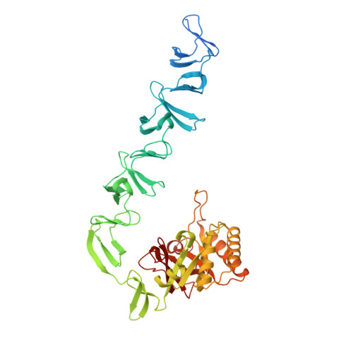
    The first structure of a pneumococcal autolysin, that of the LytC lysozyme, has been solved in ternary complex with choline and a pneumococcal peptidoglycan (PG) fragment. The active site of the hydrolase module is not fully exposed but is oriented toward the choline-binding module, which accounts for its unique in vivo features in PG hydrolysis, its activation and its regulatory mechanisms. Because of the unusual hook-shaped conformation of the multimodular protein, it is only able to hydrolyze non-cross-linked PG chains, an assertion validated by additional experiments. These results explain the activation of LytC by choline-binding protein D (CbpD) in fratricide, a competence-programmed mechanism of predation of noncompetent sister cells. The results provide the first structural insights to our knowledge into the critical and central function that LytC plays in pneumococcal virulence and explain a long-standing puzzle of how murein hydrolases can be controlled to avoid self-lysis during bacterial growth and division.


  • Organizational Affiliation

    Grupo de Cristalografía Macromolecular y Biología Estructural, Instituto Química-Física Rocasolano, Consejo Superior de Investigaciones Científicas, Madrid, Spain.


Macromolecules
Find similar proteins by:  (by identity cutoff)  |  3D Structure
Entity ID: 1
MoleculeChains Sequence LengthOrganismDetailsImage
1,4-BETA-N-ACETYLMURAMIDASE468Streptococcus pneumoniae R6Mutation(s): 0 
EC: 3.2.1.17
UniProt
Find proteins for Q8DP07 (Streptococcus pneumoniae (strain ATCC BAA-255 / R6))
Explore Q8DP07 
Go to UniProtKB:  Q8DP07
Entity Groups  
Sequence Clusters30% Identity50% Identity70% Identity90% Identity95% Identity100% Identity
UniProt GroupQ8DP07
Sequence Annotations
Expand
  • Reference Sequence
Small Molecules
Ligands 3 Unique
IDChains Name / Formula / InChI Key2D Diagram3D Interactions
CHT
Query on CHT

Download Ideal Coordinates CCD File 
I [auth A]
J [auth A]
K [auth A]
L [auth A]
M [auth A]
I [auth A],
J [auth A],
K [auth A],
L [auth A],
M [auth A],
N [auth A],
O [auth A]
CHOLINE ION
C5 H14 N O
OEYIOHPDSNJKLS-UHFFFAOYSA-N
GOL
Query on GOL

Download Ideal Coordinates CCD File 
B [auth A]
C [auth A]
D [auth A]
E [auth A]
F [auth A]
B [auth A],
C [auth A],
D [auth A],
E [auth A],
F [auth A],
G [auth A],
H [auth A]
GLYCEROL
C3 H8 O3
PEDCQBHIVMGVHV-UHFFFAOYSA-N
CL
Query on CL

Download Ideal Coordinates CCD File 
P [auth A],
Q [auth A]
CHLORIDE ION
Cl
VEXZGXHMUGYJMC-UHFFFAOYSA-M
Experimental Data & Validation

Experimental Data

  • Method: X-RAY DIFFRACTION
  • Resolution: 1.61 Å
  • R-Value Free: 0.204 
  • R-Value Work: 0.183 
  • R-Value Observed: 0.185 
  • Space Group: P 1 21 1
Unit Cell:
Length ( Å )Angle ( ˚ )
a = 59.8α = 90
b = 69.38β = 106.03
c = 75.87γ = 90
Software Package:
Software NamePurpose
MOSFLMdata reduction
SCALAdata scaling
SHELXphasing
SHARPphasing
REFMACrefinement

Structure Validation

View Full Validation Report


Entry History 

Deposition Data

Revision History  (Full details and data files)

  • Version 1.0: 2010-04-21
    Type: Initial release
  • Version 1.1: 2011-07-13
    Changes: Advisory, Version format compliance
  • Version 1.2: 2011-09-21
    Changes: Database references