Simultaneous Formation of Right- and Left-Handed Anti-Parallel Coiled-Coil Interfaces by a Coil2 Fragment of Human Lamin A.
Kapinos, L.E., Burkhard, P., Herrmann, H., Aebi, U., Strelkov, S.V.(2011) J Mol Biol 408: 135
- PubMed: 21354179
- DOI: https://doi.org/10.1016/j.jmb.2011.02.037
- Primary Citation of Related Structures:
2XV5 - PubMed Abstract:
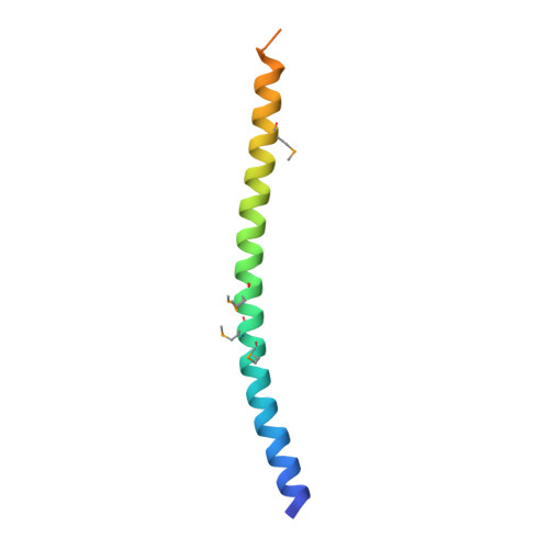
The elementary building block of all intermediate filaments (IFs) is a dimer featuring a central α-helical rod domain flanked by the N- and C-terminal end domains. In nuclear IF proteins (lamins), the rod domain consists of two coiled-coil segments, coil1 and coil2, that are connected by a short non-helical linker. Coil1 and the C-terminal part of coil2 contain the two highly conserved IF consensus motifs involved in the longitudinal assembly of dimers. The previously solved crystal structure of a lamin A fragment (residues 305-387) corresponding to the second half of coil2 has yielded a parallel left-handed coiled coil. Here, we present the crystal structure and solution properties of another human lamin A fragment (residues 328-398), which is largely overlapping with fragment 305-387 but harbors a short segment of the tail domain. Unexpectedly, no parallel coiled coil forms within the crystal. Instead, the α-helices are arranged such that two anti-parallel coiled-coil interfaces are formed. The most significant interface has a right-handed geometry, which is accounted for by a characteristic 15-residue repeat pattern that overlays with the canonical heptad repeat pattern. The second interface is a left-handed anti-parallel coiled coil based on the predicted heptad repeat pattern. In solution, the fragment reveals only a weak dimerization propensity. We speculate that the C-terminus of coil2 might unzip, thereby allowing for a right-handed coiled-coil interface to form between two laterally aligned dimers. Such an interface might co-exist with a heterotetrameric left-handed coiled-coil assembly, which is expected to be responsible for the longitudinal A(CN) contact.
Organizational Affiliation:
M.E. Müller Institute for Structural Biology, Biozentrum, University of Basel, Switzerland. larisa.kapinos@unibas.ch