2XOZ

C-terminal cysteine rich domain of human CHFR bound to AMP


Experimental Data Snapshot

  • Method: X-RAY DIFFRACTION
  • Resolution: 2.37 Å
  • R-Value Free: 0.232 
  • R-Value Work: 0.179 
  • R-Value Observed: 0.184 

wwPDB Validation   3D Report Full Report


Ligand Structure Quality Assessment 


This is version 1.2 of the entry. See complete history


Literature

Structural Basis of Poly(Adp-Ribose) Recognition by the Multizinc Binding Domain of Checkpoint with Forkhead-Associated and Ring Domains (Chfr).

Oberoi, J.Richards, M.W.Crumpler, S.Brown, N.Blagg, J.Bayliss, R.

(2010) J Biol Chem 285: 39348

  • DOI: https://doi.org/10.1074/jbc.M110.159855
  • Primary Citation of Related Structures:  
    2XOC, 2XOY, 2XOZ, 2XP0

  • PubMed Abstract: 

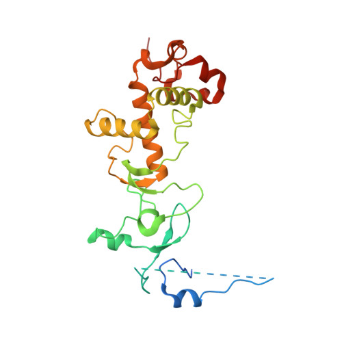
    Cellular stress in early mitosis activates the antephase checkpoint, resulting in the decondensation of chromosomes and delayed mitotic progression. Checkpoint with forkhead-associated and RING domains (CHFR) is central to this checkpoint, and its activity is ablated in many tumors and cancer cell lines through promoter hypermethylation or mutation. The interaction between the PAR-binding zinc finger (PBZ) of CHFR and poly(ADP-ribose) (PAR) is crucial for a functional antephase checkpoint. We determined the crystal structure of the cysteine-rich region of human CHFR (amino acids 425-664) to 1.9 Å resolution, which revealed a multizinc binding domain of elaborate topology within which the PBZ is embedded. The PBZ of CHFR closely resembles the analogous motifs from aprataxin-like factor and CG1218-PA, which lie within unstructured regions of their respective proteins. Based on co-crystal structures of CHFR bound to several different PAR-like ligands (adenosine 5'-diphosphoribose, adenosine monophosphate, and P(1)P(2)-diadenosine 5'-pyrophosphate), we made a model of the CHFR-PAR interaction, which we validated using site-specific mutagenesis and surface plasmon resonance. The PBZ motif of CHFR recognizes two adenine-containing subunits of PAR and the phosphate backbone that connects them. More generally, PBZ motifs may recognize different numbers of PAR subunits as required to carry out their functions.


  • Organizational Affiliation

    Section of Structural Biology, Institute of Cancer Research, Chester Beatty Laboratories, 237 Fulham Road, London SW3 6JB, United Kingdom.


Macromolecules
Find similar proteins by:  (by identity cutoff)  |  3D Structure
Entity ID: 1
MoleculeChains Sequence LengthOrganismDetailsImage
E3 UBIQUITIN-PROTEIN LIGASE CHFR
A, B
261Homo sapiensMutation(s): 0 
EC: 6.3.2
UniProt & NIH Common Fund Data Resources
Find proteins for Q96EP1 (Homo sapiens)
Explore Q96EP1 
Go to UniProtKB:  Q96EP1
PHAROS:  Q96EP1
GTEx:  ENSG00000072609 
Entity Groups  
Sequence Clusters30% Identity50% Identity70% Identity90% Identity95% Identity100% Identity
UniProt GroupQ96EP1
Sequence Annotations
Expand
  • Reference Sequence
Small Molecules
Ligands 2 Unique
IDChains Name / Formula / InChI Key2D Diagram3D Interactions
AMP
Query on AMP

Download Ideal Coordinates CCD File 
M [auth B],
N [auth B]
ADENOSINE MONOPHOSPHATE
C10 H14 N5 O7 P
UDMBCSSLTHHNCD-KQYNXXCUSA-N
ZN
Query on ZN

Download Ideal Coordinates CCD File 
C [auth A]
D [auth A]
E [auth A]
F [auth A]
G [auth A]
C [auth A],
D [auth A],
E [auth A],
F [auth A],
G [auth A],
H [auth B],
I [auth B],
J [auth B],
K [auth B],
L [auth B]
ZINC ION
Zn
PTFCDOFLOPIGGS-UHFFFAOYSA-N
Experimental Data & Validation

Experimental Data

  • Method: X-RAY DIFFRACTION
  • Resolution: 2.37 Å
  • R-Value Free: 0.232 
  • R-Value Work: 0.179 
  • R-Value Observed: 0.184 
  • Space Group: C 1 2 1
Unit Cell:
Length ( Å )Angle ( ˚ )
a = 162.81α = 90
b = 51.56β = 105.93
c = 82.49γ = 90
Software Package:
Software NamePurpose
PHENIXrefinement
MOSFLMdata reduction
SCALAdata scaling
PHASERphasing

Structure Validation

View Full Validation Report


Ligand Structure Quality Assessment 


Entry History 

Deposition Data

Revision History  (Full details and data files)

  • Version 1.0: 2010-09-29
    Type: Initial release
  • Version 1.1: 2012-03-07
    Changes: Non-polymer description, Source and taxonomy, Version format compliance
  • Version 1.2: 2023-12-20
    Changes: Data collection, Database references, Derived calculations, Other, Refinement description