2XK3

Structure of Nek2 bound to Aminopyrazine compound 35


Experimental Data Snapshot

  • Method: X-RAY DIFFRACTION
  • Resolution: 2.20 Å
  • R-Value Free: 0.238 
  • R-Value Work: 0.185 
  • R-Value Observed: 0.188 

wwPDB Validation   3D Report Full Report


Ligand Structure Quality Assessment 


This is version 1.3 of the entry. See complete history


Literature

Aminopyrazine Inhibitors Binding to an Unusual Inactive Conformation of the Mitotic Kinase Nek2: Sar and Structural Characterization.

Whelligan, D.K.Solanki, S.Taylor, D.Thomson, D.W.Cheung, K.M.Boxall, K.Mas-Droux, C.Barillari, C.Burns, S.Grummitt, C.G.Collins, I.Van Montfort, R.L.Aherne, G.W.Bayliss, R.Hoelder, S.

(2010) J Med Chem 53: 7682

  • DOI: https://doi.org/10.1021/jm1008727
  • Primary Citation of Related Structures:  
    2XK3, 2XK4, 2XK6, 2XK7, 2XK8, 2XKC, 2XKD, 2XKE, 2XKF

  • PubMed Abstract: 

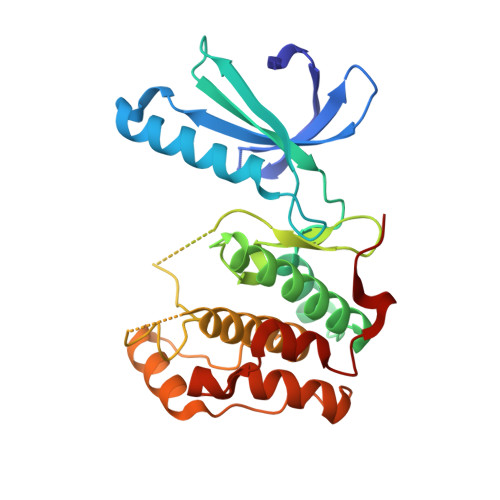
    We report herein the first systematic exploration of inhibitors of the mitotic kinase Nek2. Starting from HTS hit aminopyrazine 2, compounds with improved activity were identified using structure-based design. Our structural biology investigations reveal two notable observations. First, 2 and related compounds bind to an unusual, inactive conformation of the kinase which to the best of our knowledge has not been reported for other types of kinase inhibitors. Second, a phenylalanine residue at the center of the ATP pocket strongly affects the ability of the inhibitor to bind to the protein. The implications of these observations are discussed, and the work described here defines key features for potent and selective Nek2 inhibition, which will aid the identification of more advanced inhibitors of Nek2.


  • Organizational Affiliation

    Cancer Research UK Cancer Therapeutics Unit, The Institute of Cancer Research, 15 Cotswold Road, Sutton, Surrey SM2 5NG, United Kingdom.


Macromolecules
Find similar proteins by:  (by identity cutoff)  |  3D Structure
Entity ID: 1
MoleculeChains Sequence LengthOrganismDetailsImage
SERINE/THREONINE-PROTEIN KINASE NEK2279Homo sapiensMutation(s): 0 
EC: 2.7.11.1
UniProt & NIH Common Fund Data Resources
Find proteins for P51955 (Homo sapiens)
Explore P51955 
Go to UniProtKB:  P51955
PHAROS:  P51955
GTEx:  ENSG00000117650 
Entity Groups  
Sequence Clusters30% Identity50% Identity70% Identity90% Identity95% Identity100% Identity
UniProt GroupP51955
Sequence Annotations
Expand
  • Reference Sequence
Small Molecules
Ligands 2 Unique
IDChains Name / Formula / InChI Key2D Diagram3D Interactions
XK3
Query on XK3

Download Ideal Coordinates CCD File 
B [auth A]4-[3-AMINO-6-(3-ETHYLTHIOPHEN-2-YL)PYRAZIN-2-YL]CYCLOHEXANE-1-CARBOXYLIC ACID
C17 H21 N3 O2 S
QTTNDIPJKXHHFL-HAQNSBGRSA-N
CL
Query on CL

Download Ideal Coordinates CCD File 
C [auth A],
D [auth A]
CHLORIDE ION
Cl
VEXZGXHMUGYJMC-UHFFFAOYSA-M
Binding Affinity Annotations 
IDSourceBinding Affinity
XK3 PDBBind:  2XK3 IC50: 2100 (nM) from 1 assay(s)
Experimental Data & Validation

Experimental Data

  • Method: X-RAY DIFFRACTION
  • Resolution: 2.20 Å
  • R-Value Free: 0.238 
  • R-Value Work: 0.185 
  • R-Value Observed: 0.188 
  • Space Group: C 1 2 1
Unit Cell:
Length ( Å )Angle ( ˚ )
a = 101.03α = 90
b = 57.09β = 133.5
c = 81.41γ = 90
Software Package:
Software NamePurpose
PHENIXrefinement
MOSFLMdata reduction
SCALAdata scaling
PHASERphasing

Structure Validation

View Full Validation Report


Ligand Structure Quality Assessment 


Entry History 

Deposition Data

Revision History  (Full details and data files)

  • Version 1.0: 2010-10-27
    Type: Initial release
  • Version 1.1: 2011-05-19
    Changes: Version format compliance
  • Version 1.2: 2011-07-13
    Changes: Version format compliance
  • Version 1.3: 2023-12-20
    Changes: Data collection, Database references, Derived calculations, Other, Refinement description