2X0V

STRUCTURE OF THE P53 CORE DOMAIN MUTANT Y220C BOUND TO 4-(trifluoromethyl)benzene-1,2-diamine


Experimental Data Snapshot

  • Method: X-RAY DIFFRACTION
  • Resolution: 1.80 Å
  • R-Value Free: 0.189 
  • R-Value Work: 0.162 
  • R-Value Observed: 0.164 

wwPDB Validation   3D Report Full Report


Ligand Structure Quality Assessment 


This is version 1.4 of the entry. See complete history


Literature

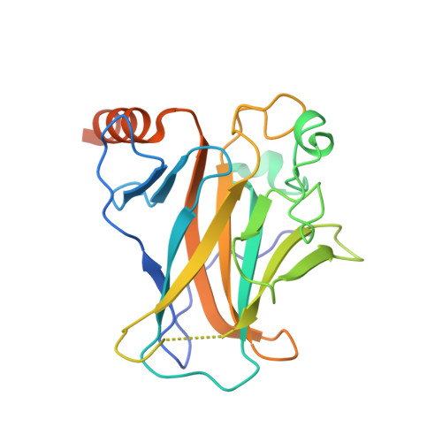
Toward the Rational Design of P53-Stabilizing Drugs: Probing the Surface of the Oncogenic Y220C Mutant.

Basse, N.Kaar, J.L.Settanni, G.Joerger, A.C.Rutherford, T.J.Fersht, A.R.

(2010) Chem Biol 17: 46

  • DOI: https://doi.org/10.1016/j.chembiol.2009.12.011
  • Primary Citation of Related Structures:  
    2X0U, 2X0V, 2X0W

  • PubMed Abstract: 

    The p53 cancer mutation Y220C induces formation of a cavity on the protein's surface that can accommodate stabilizing small molecules. We combined fragment screening and molecular dynamics to assess the druggability of p53-Y220C and map ligand interaction sites within the mutational cavity. Elucidation of the binding mode of fragment hits by crystallography yielded a clear picture of how a drug might dock in the cavity. Simulations that solvate the protein with isopropanol found additional sites that extend the druggable surface. Moreover, structural observations and simulation revealed the dynamic landscape of the cavity, which improves our understanding of the impact of the mutation on p53 stability. This underpins the importance of considering flexibility of the cavity in screening for optimized ligands. Our findings provide a blueprint for the design of effective drugs that rescue p53-Y220C.


  • Organizational Affiliation

    Medical Research Council Centre for Protein Engineering, Cambridge, UK.


Macromolecules
Find similar proteins by:  (by identity cutoff)  |  3D Structure
Entity ID: 1
MoleculeChains Sequence LengthOrganismDetailsImage
CELLULAR TUMOR ANTIGEN P53
A, B
219Homo sapiensMutation(s): 5 
UniProt & NIH Common Fund Data Resources
Find proteins for P04637 (Homo sapiens)
Explore P04637 
Go to UniProtKB:  P04637
PHAROS:  P04637
GTEx:  ENSG00000141510 
Entity Groups  
Sequence Clusters30% Identity50% Identity70% Identity90% Identity95% Identity100% Identity
UniProt GroupP04637
Sequence Annotations
Expand
  • Reference Sequence
Experimental Data & Validation

Experimental Data

  • Method: X-RAY DIFFRACTION
  • Resolution: 1.80 Å
  • R-Value Free: 0.189 
  • R-Value Work: 0.162 
  • R-Value Observed: 0.164 
  • Space Group: P 21 21 21
Unit Cell:
Length ( Å )Angle ( ˚ )
a = 65.226α = 90
b = 71.088β = 90
c = 105.389γ = 90
Software Package:
Software NamePurpose
PHENIXrefinement
PHENIXphasing

Structure Validation

View Full Validation Report


Ligand Structure Quality Assessment 


Entry History 

Deposition Data

Revision History  (Full details and data files)

  • Version 1.0: 2010-01-26
    Type: Initial release
  • Version 1.1: 2011-05-08
    Changes: Version format compliance
  • Version 1.2: 2011-07-13
    Changes: Version format compliance
  • Version 1.3: 2019-05-08
    Changes: Data collection, Experimental preparation, Other
  • Version 1.4: 2023-12-20
    Changes: Data collection, Database references, Derived calculations, Other, Refinement description