2VQJ

Structure of HDAC4 catalytic domain bound to a trifluoromethylketone inhbitor


Experimental Data Snapshot

  • Method: X-RAY DIFFRACTION
  • Resolution: 2.10 Å
  • R-Value Free: 0.276 
  • R-Value Work: 0.225 
  • R-Value Observed: 0.228 

wwPDB Validation   3D Report Full Report


Ligand Structure Quality Assessment 


This is version 1.3 of the entry. See complete history


Literature

Structural and Functional Analysis of the Human Hdac4 Catalytic Domain Reveals a Regulatory Zinc-Binding Domain.

Bottomley, M.J.Lo Surdo, P.Di Giovine, P.Cirillo, A.Scarpelli, R.Ferrigno, F.Jones, P.Neddermann, P.De Francesco, R.Steinkuhler, C.Gallinari, P.Carfi, A.

(2008) J Biol Chem 283: 26694

  • DOI: https://doi.org/10.1074/jbc.M803514200
  • Primary Citation of Related Structures:  
    2VQJ, 2VQM, 2VQO, 2VQQ, 2VQV, 2VQW

  • PubMed Abstract: 

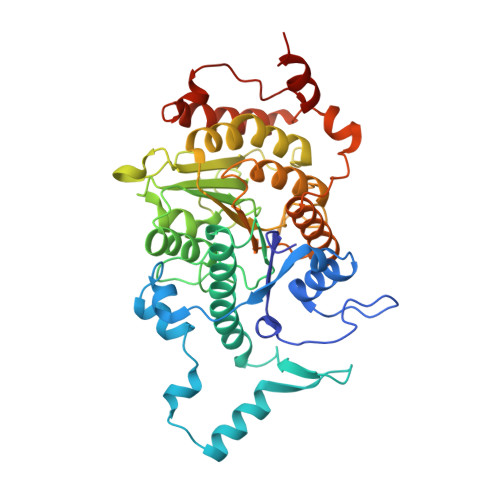
    Histone deacetylases (HDACs) regulate chromatin status and gene expression, and their inhibition is of significant therapeutic interest. To date, no biological substrate for class IIa HDACs has been identified, and only low activity on acetylated lysines has been demonstrated. Here, we describe inhibitor-bound and inhibitor-free structures of the histone deacetylase-4 catalytic domain (HDAC4cd) and of an HDAC4cd active site mutant with enhanced enzymatic activity toward acetylated lysines. The structures presented, coupled with activity data, provide the molecular basis for the intrinsically low enzymatic activity of class IIa HDACs toward acetylated lysines and reveal active site features that may guide the design of class-specific inhibitors. In addition, these structures reveal a conformationally flexible structural zinc-binding domain conserved in all class IIa enzymes. Importantly, either the mutation of residues coordinating the structural zinc ion or the binding of a class IIa selective inhibitor prevented the association of HDAC4 with the N-CoR.HDAC3 repressor complex. Together, these data suggest a key role of the structural zinc-binding domain in the regulation of class IIa HDAC functions.


  • Organizational Affiliation

    Istituto di Ricerche di Biologia Molecolare P. Angeletti, Merck Research Laboratories, Via Pontina Km 30.600, 00040 Pomezia (Roma), Italy.


Macromolecules
Find similar proteins by:  (by identity cutoff)  |  3D Structure
Entity ID: 1
MoleculeChains Sequence LengthOrganismDetailsImage
HISTONE DEACETYLASE 4413Homo sapiensMutation(s): 0 
UniProt & NIH Common Fund Data Resources
Find proteins for P56524 (Homo sapiens)
Explore P56524 
Go to UniProtKB:  P56524
PHAROS:  P56524
GTEx:  ENSG00000068024 
Entity Groups  
Sequence Clusters30% Identity50% Identity70% Identity90% Identity95% Identity100% Identity
UniProt GroupP56524
Sequence Annotations
Expand
  • Reference Sequence
Small Molecules
Ligands 5 Unique
IDChains Name / Formula / InChI Key2D Diagram3D Interactions
TFG
Query on TFG

Download Ideal Coordinates CCD File 
H [auth A]2,2,2-TRIFLUORO-1-{5-[(3-PHENYL-5,6-DIHYDROIMIDAZO[1,2-A]PYRAZIN-7(8H)-YL)CARBONYL]THIOPHEN-2-YL}ETHANE-1,1-DIOL
C19 H16 F3 N3 O3 S
OFBFUNBBOQCNFX-UHFFFAOYSA-N
SO4
Query on SO4

Download Ideal Coordinates CCD File 
E [auth A],
F [auth A],
G [auth A]
SULFATE ION
O4 S
QAOWNCQODCNURD-UHFFFAOYSA-L
DIO
Query on DIO

Download Ideal Coordinates CCD File 
B [auth A]1,4-DIETHYLENE DIOXIDE
C4 H8 O2
RYHBNJHYFVUHQT-UHFFFAOYSA-N
ZN
Query on ZN

Download Ideal Coordinates CCD File 
I [auth A],
J [auth A]
ZINC ION
Zn
PTFCDOFLOPIGGS-UHFFFAOYSA-N
K
Query on K

Download Ideal Coordinates CCD File 
C [auth A],
D [auth A]
POTASSIUM ION
K
NPYPAHLBTDXSSS-UHFFFAOYSA-N
Binding Affinity Annotations 
IDSourceBinding Affinity
TFG PDBBind:  2VQJ IC50: 367 (nM) from 1 assay(s)
Experimental Data & Validation

Experimental Data

  • Method: X-RAY DIFFRACTION
  • Resolution: 2.10 Å
  • R-Value Free: 0.276 
  • R-Value Work: 0.225 
  • R-Value Observed: 0.228 
  • Space Group: C 2 2 21
Unit Cell:
Length ( Å )Angle ( ˚ )
a = 106.77α = 90
b = 137.562β = 90
c = 68.993γ = 90
Software Package:
Software NamePurpose
REFMACrefinement
MOSFLMdata reduction
SCALAdata scaling
PHASERphasing

Structure Validation

View Full Validation Report


Ligand Structure Quality Assessment 


Entry History 

Revision History  (Full details and data files)

  • Version 1.0: 2008-07-08
    Type: Initial release
  • Version 1.1: 2011-05-08
    Changes: Version format compliance
  • Version 1.2: 2011-07-13
    Changes: Version format compliance
  • Version 1.3: 2023-12-13
    Changes: Data collection, Database references, Derived calculations, Other, Refinement description