2VEW

CRYSTAL STRUCTURE OF PROTEIN TYROSINE PHOSPHATASE 1B IN COMPLEX WITH AN ISOTHIAZOLIDINONE-CONTAINING INHIBITOR


Experimental Data Snapshot

  • Method: X-RAY DIFFRACTION
  • Resolution: 2.00 Å
  • R-Value Free: 0.239 
  • R-Value Work: 0.201 
  • R-Value Observed: 0.201 

wwPDB Validation   3D Report Full Report


Ligand Structure Quality Assessment 


This is version 1.4 of the entry. See complete history


Literature

Isothiazolidinone Inhibitors of Ptp1B Containing Imidazoles and Imidazolines

Douty, B.Wayland, B.Ala, P.J.Bower, M.J.Pruitt, J.Bostrom, L.Wei, M.Klabe, R.Gonneville, L.Wynn, R.Burn, T.C.Liu, P.C.C.Combs, A.P.Yue, E.W.

(2008) Bioorg Med Chem Lett 18: 66

  • DOI: https://doi.org/10.1016/j.bmcl.2007.11.012
  • Primary Citation of Related Structures:  
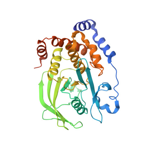
    2VEU, 2VEV, 2VEW, 2VEX, 2VEY

  • PubMed Abstract: 

    The structure-based design and synthesis of isothiazolidinone (IZD) inhibitors of PTP1B containing imidazoles and imidazolines and their modification to interact with the B site of PTP1B are described here. The X-ray crystal structures of 3I and 4I complexed with PTP1B were solved and revealed the inhibitors are interacting extensively with the B site of the enzyme.


  • Organizational Affiliation

    Incyte Corporation, Discovery Chemistry, Experimental Station, Route 141 and Henry Clay Road, Wilmington, DE 19880, USA. bdouty@incyte.com


Macromolecules
Find similar proteins by:  (by identity cutoff)  |  3D Structure
Entity ID: 1
MoleculeChains Sequence LengthOrganismDetailsImage
TYROSINE-PROTEIN PHOSPHATASE NON-RECEPTOR TYPE 1321Homo sapiensMutation(s): 0 
EC: 3.1.3.48
UniProt & NIH Common Fund Data Resources
Find proteins for P18031 (Homo sapiens)
Explore P18031 
Go to UniProtKB:  P18031
PHAROS:  P18031
GTEx:  ENSG00000196396 
Entity Groups  
Sequence Clusters30% Identity50% Identity70% Identity90% Identity95% Identity100% Identity
UniProt GroupP18031
Sequence Annotations
Expand
  • Reference Sequence
Small Molecules
Ligands 2 Unique
IDChains Name / Formula / InChI Key2D Diagram3D Interactions
IZ3
Query on IZ3

Download Ideal Coordinates CCD File 
B [auth A]3-fluoro-N-[(1S)-1-[4-[(2-fluorophenyl)methyl]imidazol-2-yl]-2-[4-[(5S)-1,1,3-trioxo-1,2-thiazolidin-5-yl]phenyl]ethyl]benzenesulfonamide
C27 H22 F2 N4 O5 S2
VAXRFGMVKHOGJO-DQEYMECFSA-N
MG
Query on MG

Download Ideal Coordinates CCD File 
C [auth A]MAGNESIUM ION
Mg
JLVVSXFLKOJNIY-UHFFFAOYSA-N
Binding Affinity Annotations 
IDSourceBinding Affinity
IZ3 PDBBind:  2VEW IC50: 64 (nM) from 1 assay(s)
Binding MOAD:  2VEW IC50: 64 (nM) from 1 assay(s)
Experimental Data & Validation

Experimental Data

  • Method: X-RAY DIFFRACTION
  • Resolution: 2.00 Å
  • R-Value Free: 0.239 
  • R-Value Work: 0.201 
  • R-Value Observed: 0.201 
  • Space Group: P 21 21 21
Unit Cell:
Length ( Å )Angle ( ˚ )
a = 66.8α = 90
b = 72.72β = 90
c = 88.13γ = 90
Software Package:
Software NamePurpose
CNSrefinement
CrystalCleardata reduction
CrystalCleardata scaling

Structure Validation

View Full Validation Report


Ligand Structure Quality Assessment 


Entry History 

Revision History  (Full details and data files)

  • Version 1.0: 2007-11-06
    Type: Initial release
  • Version 1.1: 2011-05-08
    Changes: Version format compliance
  • Version 1.2: 2011-07-13
    Changes: Version format compliance
  • Version 1.3: 2017-07-05
    Changes: Data collection
  • Version 1.4: 2020-11-18
    Changes: Derived calculations, Other, Structure summary