2V8W

Crystallographic and mass spectrometric characterisation of eIF4E with N7-cap derivatives


Experimental Data Snapshot

  • Method: X-RAY DIFFRACTION
  • Resolution: 2.30 Å
  • R-Value Free: 0.243 
  • R-Value Work: 0.184 
  • R-Value Observed: 0.188 

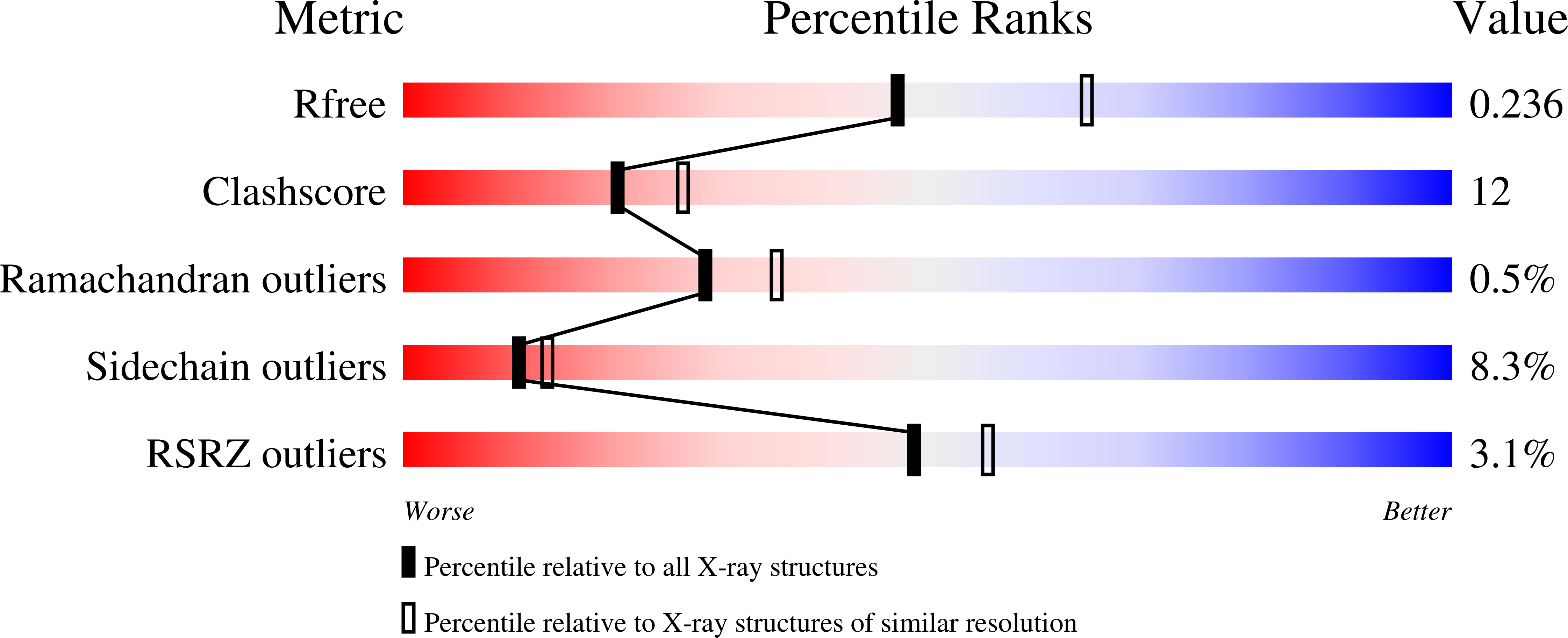
wwPDB Validation   3D Report Full Report


Ligand Structure Quality Assessment 


This is version 1.3 of the entry. See complete history


Literature

Crystallographic and Mass Spectrometric Characterisation of Eif4E with N(7)-Alkylated CAP Derivatives.

Brown, C.J.Mcnae, I.Fischer, P.M.Walkinshaw, M.D.

(2007) J Mol Biol 372: 7

  • DOI: https://doi.org/10.1016/j.jmb.2007.06.033
  • Primary Citation of Related Structures:  
    2V8W, 2V8X, 2V8Y

  • PubMed Abstract: 

    Structural complexes of the eukaryotic translation initiation factor 4E (eIF4E) with a series of N(7)-alkylated guanosine derivative mRNA cap analogue structures have been characterised. Mass spectrometry was used to determine apparent gas-phase equilibrium dissociation constants (K(d)) values of 0.15 microM, 13.6 microM, and 55.7 microM for eIF4E with 7-methyl-GTP (m(7)GTP), GTP, and GMP, respectively. For tight and specific binding to the eIF4E mononucleotide binding site, there seems to be a clear requirement for guanosine derivatives to possess both the delocalised positive charge of the N(7)-methylated guanine system and at least one phosphate group. We show that the N(7)-benzylated monophosphates 7-benzyl-GMP (Bn(7)GMP) and 7-(p-fluorobenzyl)-GMP (FBn(7)GMP) bind eIF4E substantially more tightly than non-N(7)-alkylated guanosine derivatives (K(d) values of 7.0 microM and 2.0 microM, respectively). The eIF4E complex crystal structures with Bn(7)GMP and FBn(7)GMP show that additional favourable contacts of the benzyl groups with eIF4E contribute binding energy that compensates for loss of the beta and gamma-phosphates. The N(7)-benzyl groups pack into a hydrophobic pocket behind the two tryptophan side-chains that are involved in the cation-pi stacking interaction between the cap and the eIF4E mononucleotide binding site. This pocket is formed by an induced fit in which one of the tryptophan residues involved in cap binding flips through 180 degrees relative to structures with N(7)-methylated cap derivatives. This and other observations made here will be useful in the design of new families of eIF4E inhibitors, which may have potential therapeutic applications in cancer.


  • Organizational Affiliation

    Structural Biochemistry, The University of Edinburgh, Michael Swann Building, King's Buildings, Edinburgh, EH9 3JR, Scotland, UK.


Macromolecules
Find similar proteins by:  (by identity cutoff)  |  3D Structure
Entity ID: 1
MoleculeChains Sequence LengthOrganismDetailsImage
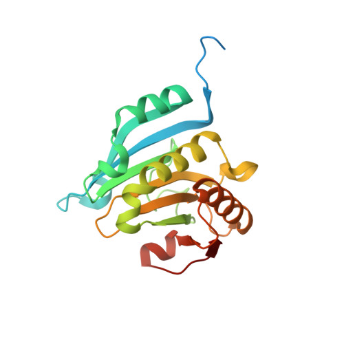
EUKARYOTIC TRANSLATION INITIATION FACTOR 4EA,
C [auth E]
217Homo sapiensMutation(s): 0 
UniProt & NIH Common Fund Data Resources
Find proteins for P06730 (Homo sapiens)
Explore P06730 
Go to UniProtKB:  P06730
PHAROS:  P06730
GTEx:  ENSG00000151247 
Entity Groups  
Sequence Clusters30% Identity50% Identity70% Identity90% Identity95% Identity100% Identity
UniProt GroupP06730
Sequence Annotations
Expand
  • Reference Sequence

Find similar proteins by:  Sequence   |   3D Structure  

Entity ID: 2
MoleculeChains Sequence LengthOrganismDetailsImage
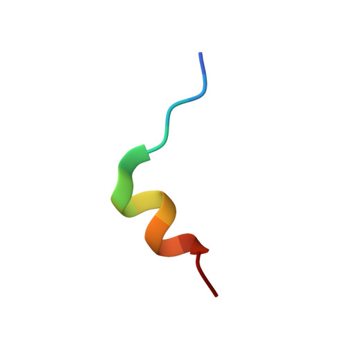
EUKARYOTIC TRANSLATION INITIATION FACTOR 4E-BINDING PROTEIN 1B,
D [auth F]
14Homo sapiensMutation(s): 0 
UniProt & NIH Common Fund Data Resources
Find proteins for Q13541 (Homo sapiens)
Explore Q13541 
Go to UniProtKB:  Q13541
PHAROS:  Q13541
GTEx:  ENSG00000187840 
Entity Groups  
Sequence Clusters30% Identity50% Identity70% Identity90% Identity95% Identity100% Identity
UniProt GroupQ13541
Sequence Annotations
Expand
  • Reference Sequence
Small Molecules
Ligands 1 Unique
IDChains Name / Formula / InChI Key2D Diagram3D Interactions
MGO
Query on MGO

Download Ideal Coordinates CCD File 
E [auth A],
F [auth E]
[[(2R,3S,4R,5R)-5-(6-AMINO-3-METHYL-4-OXO-5H-IMIDAZO[4,5-C]PYRIDIN-1-YL)-3,4-DIHYDROXY-OXOLAN-2-YL]METHOXY-HYDROXY-PHOSPHORYL] PHOSPHONO HYDROGEN PHOSPHATE
C12 H20 N4 O14 P3
LPHBJZOLBGBNJF-XYHAGOFUSA-O
Binding Affinity Annotations 
IDSourceBinding Affinity
MGO PDBBind:  2V8W Kd: 150 (nM) from 1 assay(s)
Binding MOAD:  2V8W Kd: 146 (nM) from 1 assay(s)
Experimental Data & Validation

Experimental Data

  • Method: X-RAY DIFFRACTION
  • Resolution: 2.30 Å
  • R-Value Free: 0.243 
  • R-Value Work: 0.184 
  • R-Value Observed: 0.188 
  • Space Group: P 21 21 21
Unit Cell:
Length ( Å )Angle ( ˚ )
a = 37.993α = 90
b = 99.61β = 90
c = 135.331γ = 90
Software Package:
Software NamePurpose
REFMACrefinement
MOSFLMdata reduction
SCALAdata scaling
MOLREPphasing

Structure Validation

View Full Validation Report


Ligand Structure Quality Assessment 


Entry History 

Deposition Data

Revision History  (Full details and data files)

  • Version 1.0: 2007-08-28
    Type: Initial release
  • Version 1.1: 2011-05-08
    Changes: Version format compliance
  • Version 1.2: 2011-07-13
    Changes: Version format compliance
  • Version 1.3: 2023-12-13
    Changes: Data collection, Database references, Refinement description