2V1Y

Structure of a phosphoinositide 3-kinase alpha adaptor-binding domain (ABD) in a complex with the iSH2 domain from p85 alpha


Experimental Data Snapshot

  • Method: X-RAY DIFFRACTION
  • Resolution: 2.40 Å
  • R-Value Free: 0.292 
  • R-Value Work: 0.233 
  • R-Value Observed: 0.236 

wwPDB Validation   3D Report Full Report


This is version 1.1 of the entry. See complete history


Literature

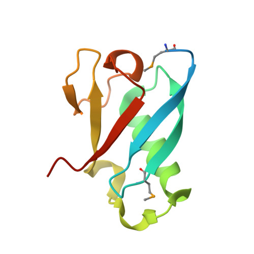
Mechanism of Two Classes of Cancer Mutations in the Phosphoinositide 3-Kinase Catalytic Subunit.

Miled, N.Yan, Y.Hon, W.C.Perisic, O.Zvelebil, M.Inbar, Y.Schneidman-Duhovny, D.Wolfson, H.J.Backer, J.M.Williams, R.L.

(2007) Science 317: 239

  • DOI: https://doi.org/10.1126/science.1135394
  • Primary Citation of Related Structures:  
    2V1Y

  • PubMed Abstract: 

    Many human cancers involve up-regulation of the phosphoinositide 3-kinase PI3Kalpha, with oncogenic mutations identified in both the p110alpha catalytic and the p85alpha regulatory subunits. We used crystallographic and biochemical approaches to gain insight into activating mutations in two noncatalytic p110alpha domains-the adaptor-binding and the helical domains. A structure of the adaptor-binding domain of p110alpha in a complex with the p85alpha inter-Src homology 2 (inter-SH2) domain shows that oncogenic mutations in the adaptor-binding domain are not at the inter-SH2 interface but in a polar surface patch that is a plausible docking site for other domains in the holo p110/p85 complex. We also examined helical domain mutations and found that the Glu545 to Lys545 (E545K) oncogenic mutant disrupts an inhibitory charge-charge interaction with the p85 N-terminal SH2 domain. These studies extend our understanding of the architecture of PI3Ks and provide insight into how two classes of mutations that cause a gain in function can lead to cancer.


  • Organizational Affiliation

    Medical Research Council Laboratory of Molecular Biology, Hills Road, Cambridge CB2 2QH, UK.


Macromolecules
Find similar proteins by:  (by identity cutoff)  |  3D Structure
Entity ID: 1
MoleculeChains Sequence LengthOrganismDetailsImage
PHOSPHATIDYLINOSITOL-4,5-BISPHOSPHATE 3-KINASE CATALYTIC SUBUNIT ALPHA ISOFORM108Bos taurusMutation(s): 0 
EC: 2.7.1.153
UniProt
Find proteins for P32871 (Bos taurus)
Explore P32871 
Go to UniProtKB:  P32871
Entity Groups  
Sequence Clusters30% Identity50% Identity70% Identity90% Identity95% Identity100% Identity
UniProt GroupP32871
Sequence Annotations
Expand
  • Reference Sequence
Find similar proteins by:  (by identity cutoff)  |  3D Structure
Entity ID: 2
MoleculeChains Sequence LengthOrganismDetailsImage
PHOSPHATIDYLINOSITOL 3-KINASE REGULATORY SUBUNIT ALPHA170Homo sapiensMutation(s): 0 
EC: 2.7.1.153
UniProt & NIH Common Fund Data Resources
Find proteins for P27986 (Homo sapiens)
Explore P27986 
Go to UniProtKB:  P27986
PHAROS:  P27986
GTEx:  ENSG00000145675 
Entity Groups  
Sequence Clusters30% Identity50% Identity70% Identity90% Identity95% Identity100% Identity
UniProt GroupP27986
Sequence Annotations
Expand
  • Reference Sequence
Small Molecules
Modified Residues  1 Unique
IDChains TypeFormula2D DiagramParent
MSE
Query on MSE
A
L-PEPTIDE LINKINGC5 H11 N O2 SeMET
Experimental Data & Validation

Experimental Data

  • Method: X-RAY DIFFRACTION
  • Resolution: 2.40 Å
  • R-Value Free: 0.292 
  • R-Value Work: 0.233 
  • R-Value Observed: 0.236 
  • Space Group: P 21 21 21
Unit Cell:
Length ( Å )Angle ( ˚ )
a = 58.799α = 90
b = 62β = 90
c = 74.654γ = 90
Software Package:
Software NamePurpose
MOSFLMdata reduction
SCALAdata scaling
SnBphasing
autoSHARPphasing
REFMACrefinement

Structure Validation

View Full Validation Report


Entry History 

Deposition Data

Revision History  (Full details and data files)

  • Version 1.0: 2007-07-24
    Type: Initial release
  • Version 1.1: 2011-07-13
    Changes: Advisory, Version format compliance