2V0O

FCHO2 F-BAR domain


Experimental Data Snapshot

  • Method: X-RAY DIFFRACTION
  • Resolution: 2.30 Å
  • R-Value Free: 0.316 
  • R-Value Work: 0.278 
  • R-Value Observed: 0.280 

wwPDB Validation   3D Report Full Report


This is version 1.2 of the entry. See complete history


Literature

Structure and Analysis of Fcho2 F-Bar Domain: A Dimerizing and Membrane Recruitment Module that Effects Membrane Curvature.

Henne, W.M.Kent, H.M.Ford, M.J.G.Hedge, B.G.Daumke, O.Butler, P.J.Mittal, R.Langen, R.Evans, P.R.Mcmahon, H.T.

(2007) Structure 15: 839

  • DOI: https://doi.org/10.1016/j.str.2007.05.002
  • Primary Citation of Related Structures:  
    2V0O

  • PubMed Abstract: 

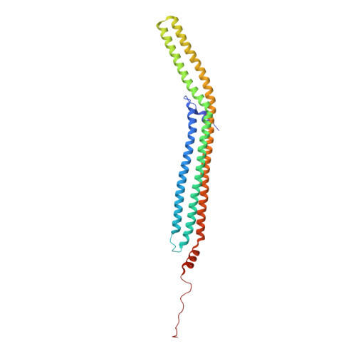
    A spectrum of membrane curvatures exists within cells, and proteins have evolved different modules to detect, create, and maintain these curvatures. Here we present the crystal structure of one such module found within human FCHo2. This F-BAR (extended FCH) module consists of two F-BAR domains, forming an intrinsically curved all-helical antiparallel dimer with a Kd of 2.5 microM. The module binds liposomes via a concave face, deforming them into tubules with variable diameters of up to 130 nm. Pulse EPR studies showed the membrane-bound dimer is the same as the crystal dimer, although the N-terminal helix changed conformation on membrane binding. Mutation of a phenylalanine on this helix partially attenuated narrow tubule formation, and resulted in a gain of curvature sensitivity. This structure shows a distant relationship to curvature-sensing BAR modules, and suggests how similar coiled-coil architectures in the BAR superfamily have evolved to expand the repertoire of membrane-sculpting possibilities.


  • Organizational Affiliation

    MRC Laboratory of Molecular Biology, Hills Road, Cambridge, CB1 0QH, United Kingdom.


Macromolecules
Find similar proteins by:  (by identity cutoff)  |  3D Structure
Entity ID: 1
MoleculeChains Sequence LengthOrganismDetailsImage
FCH DOMAIN ONLY PROTEIN 2
A, B, C
276Homo sapiensMutation(s): 0 
Membrane Entity: Yes 
UniProt & NIH Common Fund Data Resources
Find proteins for Q0JRZ9 (Homo sapiens)
Explore Q0JRZ9 
Go to UniProtKB:  Q0JRZ9
PHAROS:  Q0JRZ9
GTEx:  ENSG00000157107 
Entity Groups  
Sequence Clusters30% Identity50% Identity70% Identity90% Identity95% Identity100% Identity
UniProt GroupQ0JRZ9
Sequence Annotations
Expand
  • Reference Sequence
Experimental Data & Validation

Experimental Data

  • Method: X-RAY DIFFRACTION
  • Resolution: 2.30 Å
  • R-Value Free: 0.316 
  • R-Value Work: 0.278 
  • R-Value Observed: 0.280 
  • Space Group: C 1 2 1
Unit Cell:
Length ( Å )Angle ( ˚ )
a = 254.44α = 90
b = 65.68β = 110.32
c = 89.91γ = 90
Software Package:
Software NamePurpose
REFMACrefinement
MOSFLMdata reduction
SCALAdata scaling
SHARPphasing

Structure Validation

View Full Validation Report


Entry History 

Deposition Data

Revision History  (Full details and data files)

  • Version 1.0: 2007-06-26
    Type: Initial release
  • Version 1.1: 2011-05-08
    Changes: Version format compliance
  • Version 1.2: 2011-07-13
    Changes: Version format compliance