A catalytic mechanism that explains a low catalytic activity of serine dehydratase like-1 from human cancer cells: Crystal structure and site-directed mutagenesis studies.
Yamada, T., Komoto, J., Kasuya, T., Takata, Y., Ogawa, H., Mori, H., Takusagawa, F.(2008) Biochim Biophys Acta 1780: 809-818
- PubMed: 18342636
- DOI: https://doi.org/10.1016/j.bbagen.2008.01.020
- Primary Citation of Related Structures:
2RKB - PubMed Abstract:
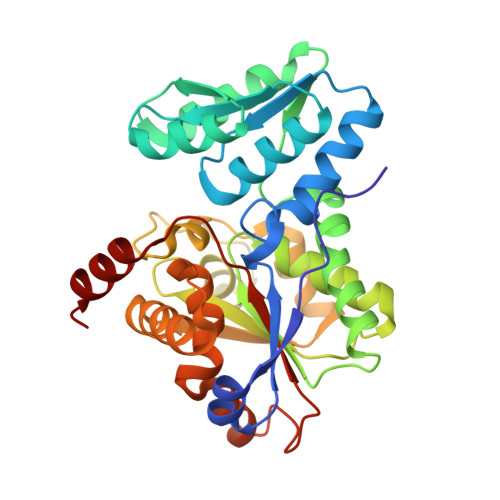
SDH (l-serine dehydratase, EC 4.3.1.17) is a pyridoxal-5'-phosphate (PLP)-dependent enzyme that catalyzes dehydration of l-Ser/Thr to yield pyruvate/ketobutyrate and ammonia. A SDH isoform (cSDH) found in human cancer cell lines has relatively low catalytic activity in comparison with the liver enzyme (hSDH). The crystal structure of cSDH has been determined at 2.8 angstroms resolution. A PLP is covalently attached to K48 by Schiff-base linkage in the active site. The ring nitrogen of PLP is involved in a H-bonding with C309, but is apparently not protonated. Twenty-three amino residues that compose the active site surfaces were identified. The human and rat liver enzymes (hSDH and rSDH) have the same residues, while residues G72, A172, and S228 in cSDH are replaced with A66, S166, and A222, respectively, in hSDH. These residues in hSDH and cSDH were mutated to make complementary pairs of mutated enzymes, and their kinetic parameters were determined. C303 of hSDH and C309 of cSDH which are H-bonding partner of the ring nitrogen of PLP were mutated to alanine and their kinetic parameters were also determined. The crystal structures and the mutation data suggest that having a glycine at residue 72 of cSDH is the major reason for the reduction of catalytic activity of cSDH. Changing alanine to glycine at residue 72 increases the flexibility of the substrate binding-loop (71S(G/A)GN74), so that the bound substrate and PLP are not pushed deep into the active cleft. Consequently, the proton transfer rate from S(G) of C309 to N1 of the bound PLP is decreased, which determines the rate of catalytic reaction.
Organizational Affiliation:
Department of Molecular Biosciences, University of Kansas, 1200 Sunnyside Avenue, Lawrence, KS 66045-7534, USA.