2QLJ

Crystal Structure of Caspase-7 with Inhibitor AC-WEHD-CHO


Experimental Data Snapshot

  • Method: X-RAY DIFFRACTION
  • Resolution: 2.60 Å
  • R-Value Free: 0.234 
  • R-Value Work: 0.196 
  • R-Value Observed: 0.201 

wwPDB Validation   3D Report Full Report


This is version 1.4 of the entry. See complete history


Literature

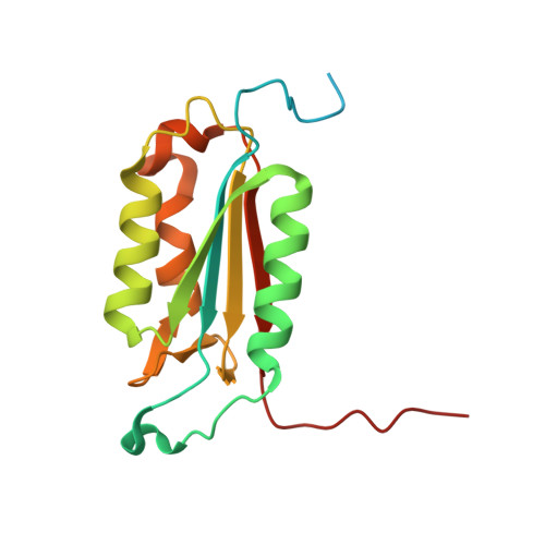
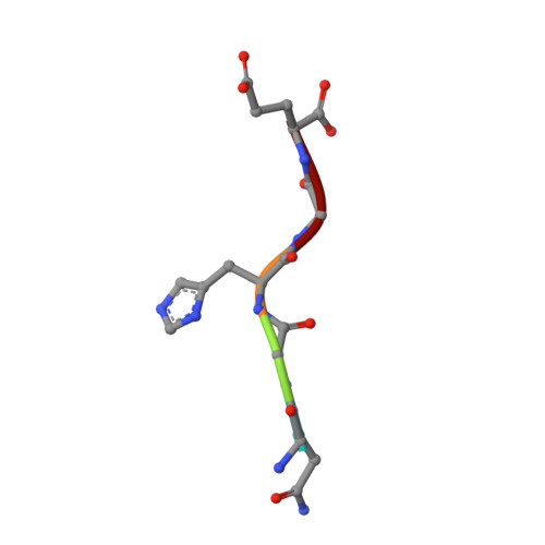
Plasticity of S2-S4 specificity pockets of executioner caspase-7 revealed by structural and kinetic analysis.

Agniswamy, J.Fang, B.Weber, I.T.

(2007) FEBS J 274: 4752-4765

  • DOI: https://doi.org/10.1111/j.1742-4658.2007.05994.x
  • Primary Citation of Related Structures:  
    2QL5, 2QL7, 2QL9, 2QLB, 2QLF, 2QLJ

  • PubMed Abstract: 

    Many protein substrates of caspases are cleaved at noncanonical sites in comparison to the recognition motifs reported for the three caspase subgroups. To provide insight into the specificity and aid in the design of drugs to control cell death, crystal structures of caspase-7 were determined in complexes with six peptide analogs (Ac-DMQD-Cho, Ac-DQMD-Cho, Ac-DNLD-Cho, Ac-IEPD-Cho, Ac-ESMD-Cho, Ac-WEHD-Cho) that span the major recognition motifs of the three subgroups. The crystal structures show that the S2 pocket of caspase-7 can accommodate diverse residues. Glu is not required at the P3 position because Ac-DMQD-Cho, Ac-DQMD-Cho and Ac-DNLD-Cho with varied P3 residues are almost as potent as the canonical Ac-DEVD-Cho. P4 Asp was present in the better inhibitors of caspase-7. However, the S4 pocket of executioner caspase-7 has alternate regions for binding of small branched aliphatic or polar residues similar to those of initiator caspase-8. The observed plasticity of the caspase subsites agrees very well with the reported cleavage of many proteins at noncanonical sites. The results imply that factors other than the P4-P1 sequence, such as exosites, contribute to the in vivo substrate specificity of caspases. The novel peptide binding site identified on the molecular surface of the current structures is suggested to be an exosite of caspase-7. These results should be considered in the design of selective small molecule inhibitors of this pharmacologically important protease.


  • Organizational Affiliation

    Department of Biology, Molecular Basis of Disease, Georgia State University, Atlanta, GA 30302, USA.


Macromolecules
Find similar proteins by:  (by identity cutoff)  |  3D Structure
Entity ID: 1
MoleculeChains Sequence LengthOrganismDetailsImage
Caspase-7
A, C
173Homo sapiensMutation(s): 0 
Gene Names: CASP7MCH3
EC: 3.4.22.60
UniProt & NIH Common Fund Data Resources
Find proteins for P55210 (Homo sapiens)
Explore P55210 
Go to UniProtKB:  P55210
PHAROS:  P55210
GTEx:  ENSG00000165806 
Entity Groups  
Sequence Clusters30% Identity50% Identity70% Identity90% Identity95% Identity100% Identity
UniProt GroupP55210
Sequence Annotations
Expand
  • Reference Sequence
Find similar proteins by:  (by identity cutoff)  |  3D Structure
Entity ID: 2
MoleculeChains Sequence LengthOrganismDetailsImage
Caspase-7
B, D
97Homo sapiensMutation(s): 0 
Gene Names: CASP7MCH3
EC: 3.4.22.60
UniProt & NIH Common Fund Data Resources
Find proteins for P55210 (Homo sapiens)
Explore P55210 
Go to UniProtKB:  P55210
PHAROS:  P55210
GTEx:  ENSG00000165806 
Entity Groups  
Sequence Clusters30% Identity50% Identity70% Identity90% Identity95% Identity100% Identity
UniProt GroupP55210
Sequence Annotations
Expand
  • Reference Sequence

Find similar proteins by:  Sequence   |   3D Structure  

Entity ID: 3
MoleculeChains Sequence LengthOrganismDetailsImage
Inhibitor AC-WEHD-CHO
E, F
5N/AMutation(s): 0 
Sequence Annotations
Expand
  • Reference Sequence

Find similar proteins by:  Sequence   |   3D Structure  

Entity ID: 4
MoleculeChains Sequence LengthOrganismDetailsImage
Peptide QGHGE5N/AMutation(s): 0 
Sequence Annotations
Expand
  • Reference Sequence
Small Molecules
Ligands 1 Unique
IDChains Name / Formula / InChI Key2D Diagram3D Interactions
CIT
Query on CIT

Download Ideal Coordinates CCD File 
H [auth A]CITRIC ACID
C6 H8 O7
KRKNYBCHXYNGOX-UHFFFAOYSA-N
Experimental Data & Validation

Experimental Data

  • Method: X-RAY DIFFRACTION
  • Resolution: 2.60 Å
  • R-Value Free: 0.234 
  • R-Value Work: 0.196 
  • R-Value Observed: 0.201 
  • Space Group: P 32 2 1
Unit Cell:
Length ( Å )Angle ( ˚ )
a = 88.504α = 90
b = 88.504β = 90
c = 187.22γ = 120
Software Package:
Software NamePurpose
HKL-2000data collection
PHASERphasing
CNSrefinement
HKL-2000data reduction
SCALEPACKdata scaling

Structure Validation

View Full Validation Report


Entry History 

Deposition Data

Revision History  (Full details and data files)

  • Version 1.0: 2007-08-28
    Type: Initial release
  • Version 1.1: 2011-07-13
    Changes: Atomic model, Database references, Derived calculations, Non-polymer description, Structure summary, Version format compliance
  • Version 1.2: 2012-12-12
    Changes: Other
  • Version 1.3: 2023-08-30
    Changes: Data collection, Database references, Derived calculations, Refinement description
  • Version 1.4: 2023-11-15
    Changes: Data collection