2PNR

Crystal Structure of the asymmetric Pdk3-l2 Complex


Experimental Data Snapshot

  • Method: X-RAY DIFFRACTION
  • Resolution: 2.50 Å
  • R-Value Free: 0.229 
  • R-Value Work: 0.176 
  • R-Value Observed: 0.185 

wwPDB Validation   3D Report Full Report


Ligand Structure Quality Assessment 


This is version 1.2 of the entry. See complete history


Literature

Crystal Structure of an Asymmetric complex of Pyruvate Dehydrogenase Kinase 3 with Lipoyl domain 2 and its Biological Implications

Devedjiev, Y.Steussy, C.N.Vassylyev, D.G.

(2007) J Mol Biol 370: 407-416

  • DOI: https://doi.org/10.1016/j.jmb.2007.04.083
  • Primary Citation of Related Structures:  
    2PNR

  • PubMed Abstract: 

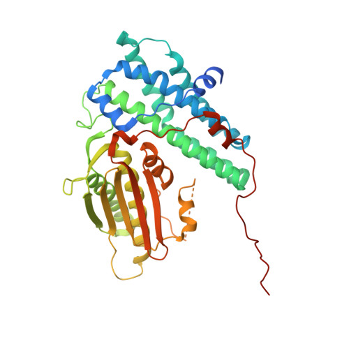
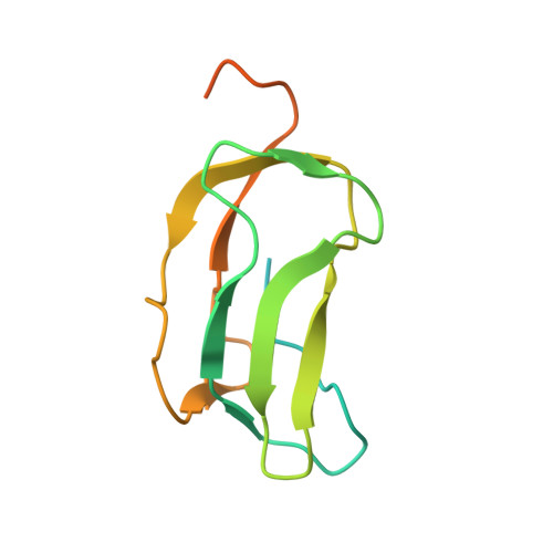
    A homodimer of pyruvate dehydrogenase kinase (PDHK) is an integral part of pyruvate dehydrogenase complex (PDC) to which it is anchored primarily through the inner lipoyl-bearing domains (L2) of transacetylase component. The catalytic cycle of PDHK and its translocation over the PDC surface is thought to be mediated by the "symmetric" and "asymmetric" modes, in which the PDHK dimer binds to two and one L2-domain(s), respectively. Whereas the structure of the symmetric PDHK/L2 complex was reported, the structural organization and functional role of the asymmetric complex remain obscure. Here, we report the crystal structure of the asymmetric PDHK3/L2 complex that reveals several functionally important features absent from the previous structures. First, the PDHK3 subunits have distinct conformations: one subunit exhibits "open" and the other "closed" configuration of the putative substrate-binding cleft. Second, access to the closed cleft is additionally restricted by local unwinding of the adjacent alpha-helix. Modeling indicates that the target peptide might gain access to the PDHK active center through the open but not through the closed cleft. Third, the ATP-binding loop in one PDHK3 subunit adopts an open conformation, implying that the nucleotide loading into the active site is mediated by the inactive "pre-insertion" binding mode. Altogether our data suggest that the asymmetric complex represents a physiological state in which binding of a single L2-domain activates one of the PDHK protomers while inactivating another. Thus, the L2-domains likely act not only as the structural anchors but also modulate the catalytic cycle of PDHK.


  • Organizational Affiliation

    Department of Biochemistry and Molecular Genetics, University of Alabama at Birmingham, School of Medicine and Dentistry, Kaul Genetics Building, Birmingham, Al 35294, USA.


Macromolecules
Find similar proteins by:  (by identity cutoff)  |  3D Structure
Entity ID: 1
MoleculeChains Sequence LengthOrganismDetailsImage
[Pyruvate dehydrogenase [lipoamide]] kinase isozyme 3A,
B,
D [auth E],
E [auth F]
419Homo sapiensMutation(s): 0 
Gene Names: PDK3
EC: 2.7.11.2
UniProt & NIH Common Fund Data Resources
Find proteins for Q15120 (Homo sapiens)
Explore Q15120 
Go to UniProtKB:  Q15120
PHAROS:  Q15120
GTEx:  ENSG00000067992 
Entity Groups  
Sequence Clusters30% Identity50% Identity70% Identity90% Identity95% Identity100% Identity
UniProt GroupQ15120
Sequence Annotations
Expand
  • Reference Sequence
Find similar proteins by:  (by identity cutoff)  |  3D Structure
Entity ID: 2
MoleculeChains Sequence LengthOrganismDetailsImage
Dihydrolipoyllysine-residue acetyltransferase component of pyruvate dehydrogenase complexC,
F [auth G]
128Homo sapiensMutation(s): 0 
Gene Names: DLATDLTA
EC: 2.3.1.12
UniProt & NIH Common Fund Data Resources
Find proteins for P10515 (Homo sapiens)
Explore P10515 
Go to UniProtKB:  P10515
PHAROS:  P10515
GTEx:  ENSG00000150768 
Entity Groups  
Sequence Clusters30% Identity50% Identity70% Identity90% Identity95% Identity100% Identity
UniProt GroupP10515
Sequence Annotations
Expand
  • Reference Sequence
Small Molecules
Experimental Data & Validation

Experimental Data

  • Method: X-RAY DIFFRACTION
  • Resolution: 2.50 Å
  • R-Value Free: 0.229 
  • R-Value Work: 0.176 
  • R-Value Observed: 0.185 
  • Space Group: P 41
Unit Cell:
Length ( Å )Angle ( ˚ )
a = 96.155α = 90
b = 96.155β = 90
c = 222.984γ = 90
Software Package:
Software NamePurpose
MAR345dtbdata collection
AMoREphasing
CNSrefinement
HKL-2000data reduction
HKL-2000data scaling

Structure Validation

View Full Validation Report


Ligand Structure Quality Assessment 


Entry History 

Deposition Data

Revision History  (Full details and data files)

  • Version 1.0: 2007-08-21
    Type: Initial release
  • Version 1.1: 2011-07-13
    Changes: Version format compliance
  • Version 1.2: 2023-08-30
    Changes: Data collection, Database references, Derived calculations, Refinement description