2PCX

Crystal structure of p53DBD(R282Q) at 1.54-angstrom Resolution


Experimental Data Snapshot

  • Method: X-RAY DIFFRACTION
  • Resolution: 1.54 Å
  • R-Value Free: 0.232 
  • R-Value Work: 0.191 
  • R-Value Observed: 0.191 

wwPDB Validation   3D Report Full Report


This is version 1.2 of the entry. See complete history


Literature

Impact of low-frequency hotspot mutation R282Q on the structure of p53 DNA-binding domain as revealed by crystallography at 1.54 angstroms resolution.

Tu, C.Tan, Y.H.Shaw, G.Zhou, Z.Bai, Y.Luo, R.Ji, X.

(2008) Acta Crystallogr D Biol Crystallogr 64: 471-477

  • DOI: https://doi.org/10.1107/S0907444908003338
  • Primary Citation of Related Structures:  
    2PCX

  • PubMed Abstract: 

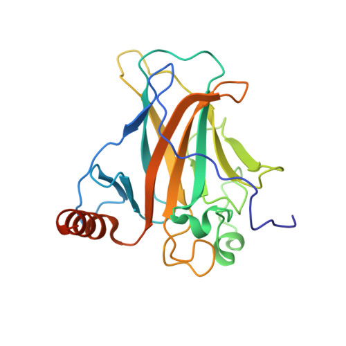
    Tumor suppressor p53 is a sequence-specific DNA-binding protein and its central DNA-binding domain (DBD) harbors six hotspots (Arg175, Gly245, Arg248, Arg249, Arg273 and Arg282) for human cancers. Here, the crystal structure of a low-frequency hotspot mutant, p53DBD(R282Q), is reported at 1.54 angstroms resolution together with the results of molecular-dynamics simulations on the basis of the structure. In addition to eliminating a salt bridge, the R282Q mutation has a significant impact on the properties of two DNA-binding loops (L1 and L3). The L1 loop is flexible in the wild type, but it is not flexible in the mutant. The L3 loop of the wild type is not flexible, whereas it assumes two conformations in the mutant. Molecular-dynamics simulations indicated that both conformations of the L3 loop are accessible under biological conditions. It is predicted that the elimination of the salt bridge and the inversion of the flexibility of L1 and L3 are directly or indirectly responsible for deactivating the tumor suppressor p53.


  • Organizational Affiliation

    Macromolecular Crystallography Laboratory, National Cancer Institute, Frederick, MD 21702, USA.


Macromolecules
Find similar proteins by:  (by identity cutoff)  |  3D Structure
Entity ID: 1
MoleculeChains Sequence LengthOrganismDetailsImage
Cellular tumor antigen p53220Homo sapiensMutation(s): 0 
Gene Names: TP53P53
UniProt & NIH Common Fund Data Resources
Find proteins for P04637 (Homo sapiens)
Explore P04637 
Go to UniProtKB:  P04637
PHAROS:  P04637
GTEx:  ENSG00000141510 
Entity Groups  
Sequence Clusters30% Identity50% Identity70% Identity90% Identity95% Identity100% Identity
UniProt GroupP04637
Sequence Annotations
Expand
  • Reference Sequence
Small Molecules
Ligands 1 Unique
IDChains Name / Formula / InChI Key2D Diagram3D Interactions
ZN
Query on ZN

Download Ideal Coordinates CCD File 
B [auth A]ZINC ION
Zn
PTFCDOFLOPIGGS-UHFFFAOYSA-N
Experimental Data & Validation

Experimental Data

  • Method: X-RAY DIFFRACTION
  • Resolution: 1.54 Å
  • R-Value Free: 0.232 
  • R-Value Work: 0.191 
  • R-Value Observed: 0.191 
  • Space Group: P 1
Unit Cell:
Length ( Å )Angle ( ˚ )
a = 34.9α = 111.8
b = 35.2β = 94.9
c = 41.6γ = 112.4
Software Package:
Software NamePurpose
CNSrefinement
HKL-2000data collection
HKL-2000data reduction
HKL-2000data scaling
AMoREphasing

Structure Validation

View Full Validation Report


Entry History 

Deposition Data

Revision History  (Full details and data files)

  • Version 1.0: 2008-04-08
    Type: Initial release
  • Version 1.1: 2011-07-13
    Changes: Version format compliance
  • Version 1.2: 2023-08-30
    Changes: Data collection, Database references, Derived calculations, Refinement description, Structure summary