2P1U

Crystal structure of the ligand binding domain of the retinoid X receptor alpha in complex with 3-(2'-ethoxy)-tetrahydronaphtyl cinnamic acid and a fragment of the coactivator TIF-2


Experimental Data Snapshot

  • Method: X-RAY DIFFRACTION
  • Resolution: 2.20 Å
  • R-Value Free: 0.251 
  • R-Value Work: 0.199 
  • R-Value Observed: 0.201 

wwPDB Validation   3D Report Full Report


Ligand Structure Quality Assessment 


This is version 1.2 of the entry. See complete history


Literature

Modulators of the structural dynamics of the retinoid X receptor to reveal receptor function.

Nahoum, V.Perez, E.Germain, P.Rodriguez-Barrios, F.Manzo, F.Kammerer, S.Lemaire, G.Hirsch, O.Royer, C.A.Gronemeyer, H.de Lera, A.R.Bourguet, W.

(2007) Proc Natl Acad Sci U S A 104: 17323-17328

  • DOI: https://doi.org/10.1073/pnas.0705356104
  • Primary Citation of Related Structures:  
    2P1T, 2P1U, 2P1V

  • PubMed Abstract: 

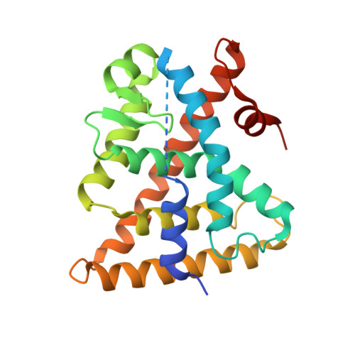
    Retinoid X receptors (RXRalpha, -beta, and -gamma) occupy a central position in the nuclear receptor superfamily, because they form heterodimers with many other family members and hence are involved in the control of a variety of (patho)physiologic processes. Selective RXR ligands, referred to as rexinoids, are already used or are being developed for cancer therapy and have promise for the treatment of metabolic diseases. However, important side effects remain associated with existing rexinoids. Here we describe the rational design and functional characterization of a spectrum of RXR modulators ranging from partial to pure antagonists and demonstrate their utility as tools to probe the implication of RXRs in cell biological phenomena. One of these ligands renders RXR activity particularly sensitive to coactivator levels and has the potential to act as a cell-specific RXR modulator. A combination of crystallographic and fluorescence anisotropy studies reveals the molecular details accounting for the agonist-to-antagonist transition and provides direct experimental evidence for a correlation between the pharmacological activity of a ligand and its impact on the structural dynamics of the activation helix H12. Using RXR and its cognate ligands as a model system, our correlative analysis of 3D structures and dynamic data provides an original view on ligand actions and enables the establishment of mechanistic concepts, which will aid in the development of selective nuclear receptor modulators.


  • Organizational Affiliation

    Institut National de la Santé et de la Recherche Médicale, U554, 34090 Montpellier, France.


Macromolecules
Find similar proteins by:  (by identity cutoff)  |  3D Structure
Entity ID: 1
MoleculeChains Sequence LengthOrganismDetailsImage
Retinoic acid receptor RXR-alpha240Homo sapiensMutation(s): 0 
Gene Names: RXRANR2B1
UniProt & NIH Common Fund Data Resources
Find proteins for P19793 (Homo sapiens)
Explore P19793 
Go to UniProtKB:  P19793
PHAROS:  P19793
GTEx:  ENSG00000186350 
Entity Groups  
Sequence Clusters30% Identity50% Identity70% Identity90% Identity95% Identity100% Identity
UniProt GroupP19793
Sequence Annotations
Expand
  • Reference Sequence

Find similar proteins by:  Sequence   |   3D Structure  

Entity ID: 2
MoleculeChains Sequence LengthOrganismDetailsImage
Nuclear receptor coactivator 2 peptide13N/AMutation(s): 0 
UniProt & NIH Common Fund Data Resources
Find proteins for Q15596 (Homo sapiens)
Explore Q15596 
Go to UniProtKB:  Q15596
PHAROS:  Q15596
GTEx:  ENSG00000140396 
Entity Groups  
Sequence Clusters30% Identity50% Identity70% Identity90% Identity95% Identity100% Identity
UniProt GroupQ15596
Sequence Annotations
Expand
  • Reference Sequence
Small Molecules
Ligands 1 Unique
IDChains Name / Formula / InChI Key2D Diagram3D Interactions
4TN
Query on 4TN

Download Ideal Coordinates CCD File 
C [auth A](2E)-3-[3-(3-ETHOXY-5,5,8,8-TETRAMETHYL-5,6,7,8-TETRAHYDRONAPHTHALEN-2-YL)-4-HYDROXYPHENYL]ACRYLIC ACID
C25 H30 O4
FXYICDGGLYXLPD-CSKARUKUSA-N
Experimental Data & Validation

Experimental Data

  • Method: X-RAY DIFFRACTION
  • Resolution: 2.20 Å
  • R-Value Free: 0.251 
  • R-Value Work: 0.199 
  • R-Value Observed: 0.201 
  • Space Group: P 43 21 2
Unit Cell:
Length ( Å )Angle ( ˚ )
a = 67.638α = 90
b = 67.638β = 90
c = 109.996γ = 90
Software Package:
Software NamePurpose
SCALAdata scaling
REFMACrefinement
PDB_EXTRACTdata extraction
DNAdata collection
MOSFLMdata reduction
MOLREPphasing

Structure Validation

View Full Validation Report


Ligand Structure Quality Assessment 


Entry History 

Deposition Data

Revision History  (Full details and data files)

  • Version 1.0: 2007-10-09
    Type: Initial release
  • Version 1.1: 2011-07-13
    Changes: Version format compliance
  • Version 1.2: 2023-08-30
    Changes: Data collection, Database references, Derived calculations, Refinement description