2OTK

Structure of Alzheimer Ab peptide in complex with an engineered binding protein


Experimental Data Snapshot

  • Method: SOLUTION NMR
  • Conformers Calculated: 100 
  • Conformers Submitted: 24 
  • Selection Criteria: structures with acceptable covalent geometry,structures with the least restraint violations,structures with the lowest energy 

wwPDB Validation   3D Report Full Report


This is version 1.2 of the entry. See complete history


Literature

Stabilization of a beta-hairpin in monomeric Alzheimer's amyloid-beta peptide inhibits amyloid formation.

Hoyer, W.Gronwall, C.Jonsson, A.Stahl, S.Hard, T.

(2008) Proc Natl Acad Sci U S A 105: 5099-5104

  • DOI: https://doi.org/10.1073/pnas.0711731105
  • Primary Citation of Related Structures:  
    2OTK

  • PubMed Abstract: 

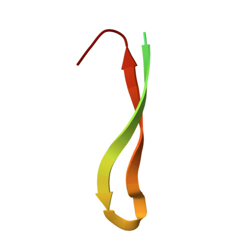
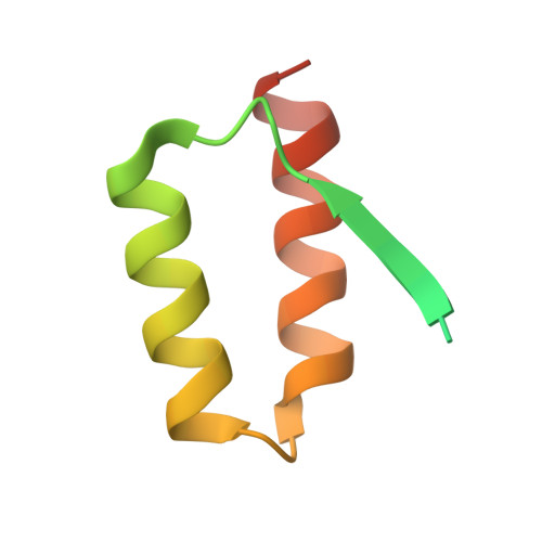
    According to the amyloid hypothesis, the pathogenesis of Alzheimer's disease is triggered by the oligomerization and aggregation of the amyloid-beta (Abeta) peptide into protein plaques. Formation of the potentially toxic oligomeric and fibrillar Abeta assemblies is accompanied by a conformational change toward a high content of beta-structure. Here, we report the solution structure of Abeta(1-40) in complex with the phage-display selected affibody protein Z(Abeta3), a binding protein of nanomolar affinity. Bound Abeta(1-40) features a beta-hairpin comprising residues 17-36, providing the first high-resolution structure of Abeta in beta conformation. The positions of the secondary structure elements strongly resemble those observed for fibrillar Abeta. Z(Abeta3) stabilizes the beta-sheet by extending it intermolecularly and by burying both of the mostly nonpolar faces of the Abeta hairpin within a large hydrophobic tunnel-like cavity. Consequently, Z(Abeta3) acts as a stoichiometric inhibitor of Abeta fibrillation. The selected Abeta conformation allows us to suggest a structural mechanism for amyloid formation based on soluble oligomeric hairpin intermediates.


  • Organizational Affiliation

    Department of Medical Biochemistry and Swedish Nuclear Magnetic Resonance Center, University of Gothenburg, Box 440, SE-405 30 Göteborg, Sweden.


Macromolecules
Find similar proteins by:  (by identity cutoff)  |  3D Structure
Entity ID: 1
MoleculeChains Sequence LengthOrganismDetailsImage
Amyloid beta A4 proteinA [auth C]40Homo sapiensMutation(s): 0 
UniProt & NIH Common Fund Data Resources
Find proteins for P05067 (Homo sapiens)
Explore P05067 
Go to UniProtKB:  P05067
PHAROS:  P05067
GTEx:  ENSG00000142192 
Entity Groups  
Sequence Clusters30% Identity50% Identity70% Identity90% Identity95% Identity100% Identity
UniProt GroupP05067
Sequence Annotations
Expand
  • Reference Sequence
Find similar proteins by:  (by identity cutoff)  |  3D Structure
Entity ID: 2
MoleculeChains Sequence LengthOrganismDetailsImage
ZAb3 Affibody dimerB [auth E],
C [auth F]
71N/AMutation(s): 0 
Entity Groups  
Sequence Clusters30% Identity50% Identity70% Identity90% Identity95% Identity100% Identity
Sequence Annotations
Expand
  • Reference Sequence
Experimental Data & Validation

Experimental Data

  • Method: SOLUTION NMR
  • Conformers Calculated: 100 
  • Conformers Submitted: 24 
  • Selection Criteria: structures with acceptable covalent geometry,structures with the least restraint violations,structures with the lowest energy 

Structure Validation

View Full Validation Report


Entry History 

Deposition Data

Revision History  (Full details and data files)

  • Version 1.0: 2008-02-12
    Type: Initial release
  • Version 1.1: 2011-07-13
    Changes: Version format compliance
  • Version 1.2: 2019-12-04
    Changes: Data collection, Derived calculations, Experimental preparation, Refinement description