2OCF

Human estrogen receptor alpha ligand-binding domain in complex with estradiol and the E2#23 FN3 monobody


Experimental Data Snapshot

  • Method: X-RAY DIFFRACTION
  • Resolution: 2.95 Å
  • R-Value Free: 0.251 
  • R-Value Work: 0.191 
  • R-Value Observed: 0.194 

wwPDB Validation   3D Report Full Report


This is version 1.5 of the entry. See complete history


Literature

Probing protein conformational changes in living cells by using designer binding proteins: application to the estrogen receptor.

Koide, A.Abbatiello, S.Rothgery, L.Koide, S.

(2002) Proc Natl Acad Sci U S A 99: 1253-1258

  • DOI: https://doi.org/10.1073/pnas.032665299
  • Primary Citation of Related Structures:  
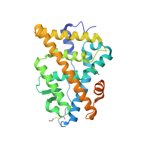
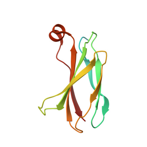
    2OCF

  • PubMed Abstract: 

    A challenge in understanding the mechanism of protein function in biology is to establish the correlation between functional form in the intracellular environment and high-resolution structures obtained with in vitro techniques. Here we present a strategy to probe conformational changes of proteins inside cells. Our method involves: (i) engineering binding proteins to different conformations of a target protein, and (ii) using them to sense changes in the surface property of the target in cells. We probed ligand-induced conformational changes of the estrogen receptor alpha (ER alpha) ligand-binding domain (LBD). By using yeast two-hybrid techniques, we first performed combinatorial library screening of "monobodies" (small antibody mimics using the scaffold of a fibronectin type III domain) for clones that bind to ER alpha and then characterized their interactions with ER alpha in the nucleus, the native environment of ER alpha, in the presence of various ligands. A library using a highly flexible loop yielded monobodies that specifically recognize a particular ligand complex of ER alpha, and the pattern of monobody specificity was consistent with the structural differences found in known crystal structures of ER alpha-LBD. A more restrained loop library yielded clones that bind both agonist- and antagonist-bound ER alpha. Furthermore, we found that a deletion of the ER alpha F domain that is C-terminally adjacent to the LBD increased the crossreactivity of monobodies to the apo-ER alpha-LBD, suggesting a dynamic nature of the ER alpha-LBD conformation and a role of the F domain in restraining the LBD in an inactive conformation.


  • Organizational Affiliation

    Department of Biochemistry and Biophysics, University of Rochester School of Medicine and Dentistry, 601 Elmwood Avenue, Rochester, NY 14642, USA.


Macromolecules
Find similar proteins by:  (by identity cutoff)  |  3D Structure
Entity ID: 1
MoleculeChains Sequence LengthOrganismDetailsImage
Estrogen receptor298Homo sapiensMutation(s): 3 
Gene Names: ESR1ESRNR3A1
UniProt & NIH Common Fund Data Resources
Find proteins for P03372 (Homo sapiens)
Explore P03372 
Go to UniProtKB:  P03372
PHAROS:  P03372
GTEx:  ENSG00000091831 
Entity Groups  
Sequence Clusters30% Identity50% Identity70% Identity90% Identity95% Identity100% Identity
UniProt GroupP03372
Sequence Annotations
Expand
  • Reference Sequence
Find similar proteins by:  (by identity cutoff)  |  3D Structure
Entity ID: 2
MoleculeChains Sequence LengthOrganismDetailsImage
FibronectinB [auth D]121Homo sapiensMutation(s): 8 
Gene Names: CIGFN1FN
UniProt & NIH Common Fund Data Resources
Find proteins for P02751 (Homo sapiens)
Explore P02751 
Go to UniProtKB:  P02751
PHAROS:  P02751
GTEx:  ENSG00000115414 
Entity Groups  
Sequence Clusters30% Identity50% Identity70% Identity90% Identity95% Identity100% Identity
UniProt GroupP02751
Sequence Annotations
Expand
  • Reference Sequence
Small Molecules
Ligands 1 Unique
IDChains Name / Formula / InChI Key2D Diagram3D Interactions
EST
Query on EST

Download Ideal Coordinates CCD File 
C [auth A]ESTRADIOL
C18 H24 O2
VOXZDWNPVJITMN-ZBRFXRBCSA-N
Modified Residues  1 Unique
IDChains TypeFormula2D DiagramParent
CME
Query on CME
A
L-PEPTIDE LINKINGC5 H11 N O3 S2CYS
Binding Affinity Annotations 
IDSourceBinding Affinity
EST BindingDB:  2OCF Ki: min: 0.11, max: 100 (nM) from 13 assay(s)
Kd: min: 0.2, max: 100 (nM) from 4 assay(s)
IC50: min: 1.00e-2, max: 46 (nM) from 46 assay(s)
EC50: min: 4.00e-3, max: 10 (nM) from 52 assay(s)
Experimental Data & Validation

Experimental Data

  • Method: X-RAY DIFFRACTION
  • Resolution: 2.95 Å
  • R-Value Free: 0.251 
  • R-Value Work: 0.191 
  • R-Value Observed: 0.194 
  • Space Group: P 61 2 2
Unit Cell:
Length ( Å )Angle ( ˚ )
a = 119.219α = 90
b = 119.219β = 90
c = 132.999γ = 120
Software Package:
Software NamePurpose
REFMACrefinement
HKL-2000data collection
HKL-2000data reduction
HKL-2000data scaling
XFITdata reduction

Structure Validation

View Full Validation Report


Entry History 

Deposition Data

Revision History  (Full details and data files)

  • Version 1.0: 2007-11-06
    Type: Initial release
  • Version 1.1: 2011-07-13
    Changes: Advisory, Version format compliance
  • Version 1.2: 2011-08-03
    Changes: Database references
  • Version 1.3: 2017-10-11
    Changes: Data collection
  • Version 1.4: 2021-10-20
    Changes: Database references, Derived calculations
  • Version 1.5: 2023-08-30
    Changes: Data collection, Refinement description