2NPQ

A Novel Lipid Binding Site in the p38 alpha MAP Kinase


Experimental Data Snapshot

  • Method: X-RAY DIFFRACTION
  • Resolution: 1.80 Å
  • R-Value Free: 0.245 
  • R-Value Work: 0.203 
  • R-Value Observed: 0.205 

wwPDB Validation   3D Report Full Report


This is version 1.3 of the entry. See complete history


Literature

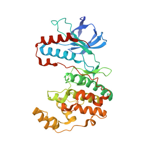
A novel lipid binding site formed by the MAP kinase insert in p38 alpha.

Diskin, R.Engelberg, D.Livnah, O.

(2008) J Mol Biol 375: 70-79

  • DOI: https://doi.org/10.1016/j.jmb.2007.09.002
  • Primary Citation of Related Structures:  
    2NPQ

  • PubMed Abstract: 

    The p38 mitogen-activated protein (MAP) kinases function as signaling molecules essential for many cellular processes, particularly mediating stress response. The activity of p38 MAP kinases is meticulously regulated to reach the desired cellular phenotype. Several alternative activation and attenuation mechanisms have been characterized recently which include new phosphorylation sites. Here we present the crystal structure of p38 alpha MAP kinase in complex with n-octyl-beta-glucopyranoside detergent. The complex unveils a novel lipid-binding site formed by a local conformational change of the MAP kinase insert. This binding is the first attribution for a possible role of the MAP kinase insert in p38. The binding site can accommodate a large selection of lipidic molecules. In addition, we also show via biophysical methods that arachidonic acid and its derivatives bind p38 alpha in vitro. Based on our analysis we propose that the binding of lipids could fine-tune p38 alpha catalytic activity towards a preferred phenotype.


  • Organizational Affiliation

    The Wolfson Centre for Applied Structural Biology, The Silberman Institute of Life Sciences, The Hebrew University of Jerusalem, Givat Ram, Jerusalem 91904, Israel.


Macromolecules
Find similar proteins by:  (by identity cutoff)  |  3D Structure
Entity ID: 1
MoleculeChains Sequence LengthOrganismDetailsImage
Mitogen-activated protein kinase 14367Homo sapiensMutation(s): 0 
EC: 2.7.11.24
UniProt & NIH Common Fund Data Resources
Find proteins for Q16539 (Homo sapiens)
Explore Q16539 
Go to UniProtKB:  Q16539
PHAROS:  Q16539
GTEx:  ENSG00000112062 
Entity Groups  
Sequence Clusters30% Identity50% Identity70% Identity90% Identity95% Identity100% Identity
UniProt GroupQ16539
Sequence Annotations
Expand
  • Reference Sequence
Small Molecules
Ligands 1 Unique
IDChains Name / Formula / InChI Key2D Diagram3D Interactions
BOG
Query on BOG

Download Ideal Coordinates CCD File 
B [auth A],
C [auth A]
octyl beta-D-glucopyranoside
C14 H28 O6
HEGSGKPQLMEBJL-RKQHYHRCSA-N
Binding Affinity Annotations 
IDSourceBinding Affinity
BOG Binding MOAD:  2NPQ Kd: 3080 (nM) from 1 assay(s)
Experimental Data & Validation

Experimental Data

  • Method: X-RAY DIFFRACTION
  • Resolution: 1.80 Å
  • R-Value Free: 0.245 
  • R-Value Work: 0.203 
  • R-Value Observed: 0.205 
  • Space Group: P 21 21 21
Unit Cell:
Length ( Å )Angle ( ˚ )
a = 68.607α = 90
b = 74.212β = 90
c = 74.571γ = 90
Software Package:
Software NamePurpose
REFMACrefinement
HKL-2000data collection
HKL-2000data reduction
HKL-2000data scaling

Structure Validation

View Full Validation Report


Entry History 

Deposition Data

Revision History  (Full details and data files)

  • Version 1.0: 2007-10-16
    Type: Initial release
  • Version 1.1: 2011-07-13
    Changes: Version format compliance
  • Version 1.2: 2020-07-29
    Type: Remediation
    Reason: Carbohydrate remediation
    Changes: Advisory, Data collection, Database references, Derived calculations, Structure summary
  • Version 1.3: 2023-10-25
    Changes: Advisory, Data collection, Database references, Refinement description, Structure summary