Molecular structural basis for polymorphism in Alzheimer's beta-amyloid fibrils.
Paravastu, A.K., Leapman, R.D., Yau, W.M., Tycko, R.(2008) Proc Natl Acad Sci U S A 105: 18349-18354
- PubMed: 19015532
- DOI: https://doi.org/10.1073/pnas.0806270105
- Primary Citation of Related Structures:
2LMN, 2LMO, 2LMP, 2LMQ - PubMed Abstract:
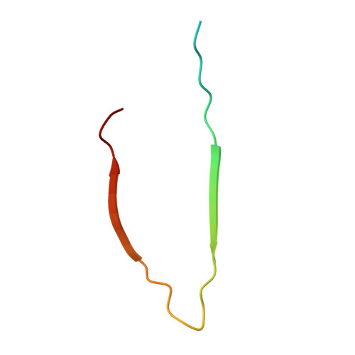
We describe a full structural model for amyloid fibrils formed by the 40-residue beta-amyloid peptide associated with Alzheimer's disease (Abeta(1-40)), based on numerous constraints from solid state NMR and electron microscopy. This model applies specifically to fibrils with a periodically twisted morphology, with twist period equal to 120 +/- 20 nm (defined as the distance between apparent minima in fibril width in negatively stained transmission electron microscope images). The structure has threefold symmetry about the fibril growth axis, implied by mass-per-length data and the observation of a single set of (13)C NMR signals. Comparison with a previously reported model for Abeta(1-40) fibrils with a qualitatively different, striated ribbon morphology reveals the molecular basis for polymorphism. At the molecular level, the 2 Abeta(1-40) fibril morphologies differ in overall symmetry (twofold vs. threefold), the conformation of non-beta-strand segments, and certain quaternary contacts. Both morphologies contain in-register parallel beta-sheets, constructed from nearly the same beta-strand segments. Because twisted and striated ribbon morphologies are also observed for amyloid fibrils formed by other polypeptides, such as the amylin peptide associated with type 2 diabetes, these structural variations may have general implications.
Organizational Affiliation:
Laboratory of Chemical Physics, National Institute of Diabetes and Digestive and Kidney Diseases, National Institutes of Health, Bethesda, MD 20892-0520, USA.