2KNE

Calmodulin wraps around its binding domain in the plasma membrane CA2+ pump anchored by a novel 18-1 motif


Experimental Data Snapshot

  • Method: SOLUTION NMR
  • Conformers Calculated: 100 
  • Conformers Submitted: 20 
  • Selection Criteria: structures with the lowest energy 

wwPDB Validation   3D Report Full Report


This is version 1.3 of the entry. See complete history


Literature

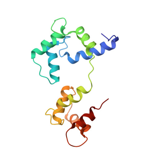
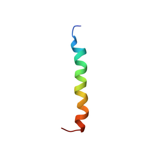
Calmodulin wraps around its binding domain in the plasma membrane Ca2+ pump anchored by a novel 18-1 motif.

Juranic, N.Atanasova, E.Filoteo, A.G.Macura, S.Prendergast, F.G.Penniston, J.T.Strehler, E.E.

(2010) J Biol Chem 285: 4015-4024

  • DOI: https://doi.org/10.1074/jbc.M109.060491
  • Primary Citation of Related Structures:  
    2KNE

  • PubMed Abstract: 

    Using solution NMR spectroscopy, we obtained the structure of Ca(2+)-calmodulin (holoCaM) in complex with peptide C28 from the binding domain of the plasma membrane Ca(2+)-ATPase (PMCA) pump isoform 4b. This provides the first atomic resolution insight into the binding mode of holoCaM to the full-length binding domain of PMCA. Structural comparison of the previously determined holoCaM.C20 complex with this holoCaM.C28 complex supports the idea that the initial binding step is represented by (holoCaM.C20) and the final bound complex by (holoCaM.C28). This affirms the existing multi-step kinetic model of PMCA4b activation by CaM. The complex exhibits a new binding motif in which holoCaM is wrapped around helical C28 peptide using two anchoring residues from the peptide at relative positions 18 and 1. The anchors correspond to Phe-1110 and Trp-1093, respectively, in full-length PMCA4b, and the peptide and CaM are oriented in an anti-parallel manner. This is a greater sequence distance between anchors than in any of the known holoCaM complexes with a helical peptide. Analysis of the geometry of holoCaM-peptide binding for the cases where the target peptide adopts an alpha(D)-helix with its anchors buried in the main hydrophobic pockets of the two CaM lobes establishes that only relative sequential positions of 10, 14, 17, and 18 are allowed for the second anchor.


  • Organizational Affiliation

    From the Departments of Biochemistry and Molecular Biology, Mayo Clinic College of Medicine, Rochester, Minnesota 55905. Electronic address: juranic.nenad@mayo.edu.


Macromolecules
Find similar proteins by:  (by identity cutoff)  |  3D Structure
Entity ID: 1
MoleculeChains Sequence LengthOrganismDetailsImage
Calmodulin148Homo sapiensMutation(s): 0 
Gene Names: 
CALMCALM1CALM2CALM3CALML2CAMCAM1CAM2CAM3CAMB...
CALMCALM1CALM2CALM3CALML2CAMCAM1CAM2CAM3CAMBCAMCCAMIII

UniProt & NIH Common Fund Data Resources
Find proteins for P0DP23 (Homo sapiens)
Explore P0DP23 
Go to UniProtKB:  P0DP23
PHAROS:  P0DP23
GTEx:  ENSG00000198668 
Entity Groups  
Sequence Clusters30% Identity50% Identity70% Identity90% Identity95% Identity100% Identity
UniProt GroupP0DP23
Sequence Annotations
Expand
  • Reference Sequence
Find similar proteins by:  (by identity cutoff)  |  3D Structure
Entity ID: 2
MoleculeChains Sequence LengthOrganismDetailsImage
ATPase, Ca++ transporting, plasma membrane 428Homo sapiensMutation(s): 0 
Gene Names: ATP2B4hCG_18445RP11-397P13.1-001
UniProt & NIH Common Fund Data Resources
Find proteins for P23634 (Homo sapiens)
Explore P23634 
Go to UniProtKB:  P23634
PHAROS:  P23634
GTEx:  ENSG00000058668 
Entity Groups  
Sequence Clusters30% Identity50% Identity70% Identity90% Identity95% Identity100% Identity
UniProt GroupP23634
Sequence Annotations
Expand
  • Reference Sequence
Experimental Data & Validation

Experimental Data

  • Method: SOLUTION NMR
  • Conformers Calculated: 100 
  • Conformers Submitted: 20 
  • Selection Criteria: structures with the lowest energy 

Structure Validation

View Full Validation Report


Entry History 

Deposition Data

Revision History  (Full details and data files)

  • Version 1.0: 2009-11-24
    Type: Initial release
  • Version 1.1: 2011-07-13
    Changes: Version format compliance
  • Version 1.2: 2020-02-26
    Changes: Data collection, Derived calculations, Other
  • Version 1.3: 2023-06-14
    Changes: Database references, Derived calculations, Other