2JGU

crystal structure of DNA-directed DNA polymerase


Experimental Data Snapshot

  • Method: X-RAY DIFFRACTION
  • Resolution: 2.60 Å
  • R-Value Free: 0.271 
  • R-Value Work: 0.275 
  • R-Value Observed: 0.275 

wwPDB Validation   3D Report Full Report


This is version 1.3 of the entry. See complete history


Literature

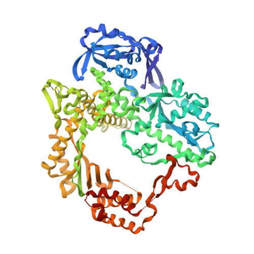
Crystal Structure of Pfu, the High Fidelity DNA Polymerase from Pyrococcus Furiosus.

Kim, S.W.Kim, D.U.Kim, J.K.Kang, L.W.Cho, H.S.

(2008) Int J Biol Macromol 42: 356

  • DOI: https://doi.org/10.1016/j.ijbiomac.2008.01.010
  • Primary Citation of Related Structures:  
    2JGU

  • PubMed Abstract: 

    We have determined a 2.6A resolution crystal structure of Pfu DNA polymerase, the most commonly used high fidelity PCR enzyme, from Pyrococcus furiosus. Although the structures of Pfu and KOD1 are highly similar, the structure of Pfu elucidates the electron density of the interface between the exonuclease and thumb domains, which has not been previously observed in the KOD1 structure. The interaction of these two domains is known to coordinate the proofreading and polymerization activity of DNA polymerases, especially via H147 that is present within the loop (residues 144-158) of the exonuclease domain. In our structure of Pfu, however, E148 rather than H147 is located at better position to interact with the thumb domain. In addition, the structural analysis of Pfu and KOD1 shows that both the Y-GG/A and beta-hairpin motifs of Pfu are found to differ with that of KOD1, and may explain differences in processivity. This information enables us to better understand the mechanisms of polymerization and proofreading of DNA polymerases.


  • Organizational Affiliation

    Department of Clinical Laboratory Science, College of Health Sciences, Korea University, Seoul 136-703, Republic of Korea.


Macromolecules
Find similar proteins by:  (by identity cutoff)  |  3D Structure
Entity ID: 1
MoleculeChains Sequence LengthOrganismDetailsImage
DNA POLYMERASE775Pyrococcus furiosusMutation(s): 0 
EC: 2.7.7.7
UniProt
Find proteins for P61875 (Pyrococcus furiosus (strain ATCC 43587 / DSM 3638 / JCM 8422 / Vc1))
Explore P61875 
Go to UniProtKB:  P61875
Entity Groups  
Sequence Clusters30% Identity50% Identity70% Identity90% Identity95% Identity100% Identity
UniProt GroupP61875
Sequence Annotations
Expand
  • Reference Sequence
Experimental Data & Validation

Experimental Data

  • Method: X-RAY DIFFRACTION
  • Resolution: 2.60 Å
  • R-Value Free: 0.271 
  • R-Value Work: 0.275 
  • R-Value Observed: 0.275 
  • Space Group: C 1 2 1
Unit Cell:
Length ( Å )Angle ( ˚ )
a = 92.178α = 90
b = 127.923β = 109.11
c = 89.21γ = 90
Software Package:
Software NamePurpose
CNSrefinement
HKL-2000data scaling
MOLREPphasing

Structure Validation

View Full Validation Report


Entry History 

Deposition Data

Revision History  (Full details and data files)

  • Version 1.0: 2008-04-22
    Type: Initial release
  • Version 1.1: 2011-05-08
    Changes: Version format compliance
  • Version 1.2: 2011-07-13
    Changes: Version format compliance
  • Version 1.3: 2023-12-13
    Changes: Data collection, Database references, Derived calculations, Other, Refinement description