2J5E

Crystal structure of EGFR kinase domain in complex with an irreversible inhibitor 13-jab


Experimental Data Snapshot

  • Method: X-RAY DIFFRACTION
  • Resolution: 3.10 Å
  • R-Value Free: 0.255 
  • R-Value Work: 0.186 
  • R-Value Observed: 0.191 

wwPDB Validation   3D Report Full Report


This is version 1.3 of the entry. See complete history


Literature

Structure-Guided Development of Affinity Probes for Tyrosine Kinases Using Chemical Genetics.

Blair, J.A.Rauh, D.Kung, C.Yun, C.-H.Fan, Q.-W.Rode, H.Zhang, C.Eck, M.J.Weiss, W.A.Shokat, K.M.

(2007) Nat Chem Biol 3: 229

  • DOI: https://doi.org/10.1038/nchembio866
  • Primary Citation of Related Structures:  
    2HWO, 2HWP, 2J5E, 2J5F

  • PubMed Abstract: 

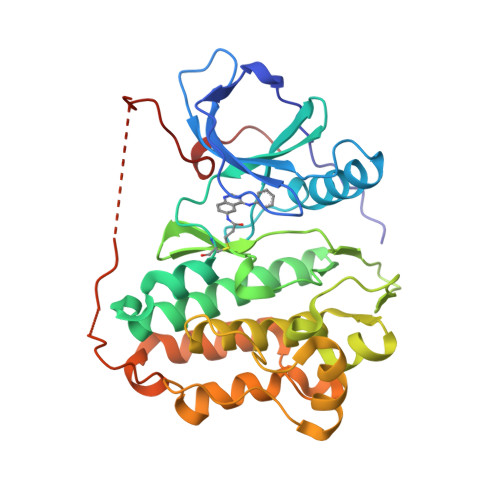
    As key components in nearly every signal transduction pathway, protein kinases are attractive targets for the regulation of cellular signaling by small-molecule inhibitors. We report the structure-guided development of 6-acrylamido-4-anilinoquinazoline irreversible kinase inhibitors that potently and selectively target rationally designed kinases bearing two selectivity elements that are not found together in any wild-type kinase: an electrophile-targeted cysteine residue and a glycine gatekeeper residue. Cocrystal structures of two irreversible quinazoline inhibitors bound to either epidermal growth factor receptor (EGFR) or engineered c-Src show covalent inhibitor binding to the targeted cysteine (Cys797 in EGFR and Cys345 in engineered c-Src). To accommodate the new covalent bond, the quinazoline core adopts positions that are different from those seen in kinase structures with reversible quinazoline inhibitors. Based on these structures, we developed a fluorescent 6-acrylamido-4-anilinoquinazoline affinity probe to report the fraction of kinase necessary for cellular signaling, and we used these reagents to quantitate the relationship between EGFR stimulation by EGF and its downstream outputs-Akt, Erk1 and Erk2.


  • Organizational Affiliation

    Department of Chemistry, University of California, Berkeley, Berkeley, California 94720, USA.


Macromolecules
Find similar proteins by:  (by identity cutoff)  |  3D Structure
Entity ID: 1
MoleculeChains Sequence LengthOrganismDetailsImage
EPIDERMAL GROWTH FACTOR RECEPTOR327Homo sapiensMutation(s): 0 
EC: 2.7.10.1
UniProt & NIH Common Fund Data Resources
Find proteins for P00533 (Homo sapiens)
Explore P00533 
Go to UniProtKB:  P00533
PHAROS:  P00533
GTEx:  ENSG00000146648 
Entity Groups  
Sequence Clusters30% Identity50% Identity70% Identity90% Identity95% Identity100% Identity
UniProt GroupP00533
Sequence Annotations
Expand
  • Reference Sequence
Small Molecules
Ligands 1 Unique
IDChains Name / Formula / InChI Key2D Diagram3D Interactions
CL
Query on CL

Download Ideal Coordinates CCD File 
B [auth A]CHLORIDE ION
Cl
VEXZGXHMUGYJMC-UHFFFAOYSA-M
Modified Residues  1 Unique
IDChains TypeFormula2D DiagramParent
CY0
Query on CY0
A
L-PEPTIDE LINKINGC20 H21 N5 O3 SCYS
Experimental Data & Validation

Experimental Data

  • Method: X-RAY DIFFRACTION
  • Resolution: 3.10 Å
  • R-Value Free: 0.255 
  • R-Value Work: 0.186 
  • R-Value Observed: 0.191 
  • Space Group: I 2 3
Unit Cell:
Length ( Å )Angle ( ˚ )
a = 145.021α = 90
b = 145.021β = 90
c = 145.021γ = 90
Software Package:
Software NamePurpose
REFMACrefinement
DENZOdata reduction
SCALEPACKdata scaling
PHASERphasing

Structure Validation

View Full Validation Report


Entry History 

Deposition Data

Revision History  (Full details and data files)

  • Version 1.0: 2007-02-27
    Type: Initial release
  • Version 1.1: 2011-05-08
    Changes: Version format compliance
  • Version 1.2: 2011-07-13
    Changes: Version format compliance
  • Version 1.3: 2023-12-13
    Changes: Data collection, Database references, Derived calculations, Other, Refinement description