2J25

Partially deglycosylated glucoceramidase


Experimental Data Snapshot

  • Method: X-RAY DIFFRACTION
  • Resolution: 2.90 Å
  • R-Value Free: 0.273 
  • R-Value Work: 0.215 
  • R-Value Observed: 0.218 

wwPDB Validation   3D Report Full Report


Ligand Structure Quality Assessment 


This is version 2.1 of the entry. See complete history


Literature

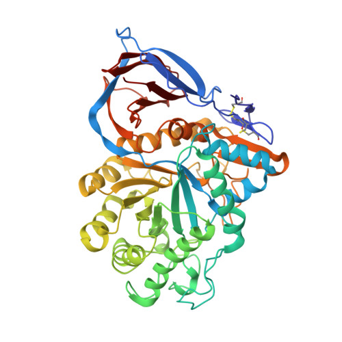
Structural Comparison of Differently Glycosylated Forms of Acid-Beta-Glucosidase, the Defective Enzyme in Gaucher Disease

Brumshtein, B.Wormald, M.R.Silman, I.Futerman, A.H.Sussman, J.L.

(2006) Acta Crystallogr D Biol Crystallogr 62: 1458

  • DOI: https://doi.org/10.1107/S0907444906038303
  • Primary Citation of Related Structures:  
    2J25

  • PubMed Abstract: 

    Gaucher disease is caused by mutations in the gene encoding acid-beta-glucosidase. A recombinant form of this enzyme, Cerezyme, is used to treat Gaucher disease patients by ;enzyme-replacement therapy'. Crystals of Cerezyme after its partial deglycosylation were obtained earlier and the structure was solved to 2.0 A resolution [Dvir et al. (2003), EMBO Rep. 4, 704-709]. The crystal structure of unmodified Cerezyme is now reported, in which a substantial number of sugar residues bound to three asparagines via N-glycosylation could be visualized. The structure of intact fully glycosylated Cerezyme is virtually identical to that of the partially deglycosylated enzyme. However, the three loops at the entrance to the active site, which were previously observed in alternative conformations, display additional variability in their structures. Comparison of the structure of acid-beta-glucosidase with that of xylanase, a bacterial enzyme from a closely related protein family, demonstrates a close correspondence between the active-site residues of the two enzymes.


  • Organizational Affiliation

    Department of Structural Biology, Weizmann Institute of Science, Israel.


Macromolecules
Find similar proteins by:  (by identity cutoff)  |  3D Structure
Entity ID: 1
MoleculeChains Sequence LengthOrganismDetailsImage
GLUCOSYLCERAMIDASE
A, B
497Homo sapiensMutation(s): 0 
EC: 3.2.1.45
UniProt & NIH Common Fund Data Resources
Find proteins for P04062 (Homo sapiens)
Explore P04062 
Go to UniProtKB:  P04062
PHAROS:  P04062
GTEx:  ENSG00000177628 
Entity Groups  
Sequence Clusters30% Identity50% Identity70% Identity90% Identity95% Identity100% Identity
UniProt GroupP04062
Sequence Annotations
Expand
  • Reference Sequence
Oligosaccharides

Help

Entity ID: 2
MoleculeChains Length2D Diagram Glycosylation3D Interactions
2-acetamido-2-deoxy-beta-D-glucopyranose-(1-4)-2-acetamido-2-deoxy-beta-D-glucopyranose
C, F
2N-Glycosylation
Glycosylation Resources
GlyTouCan:  G42666HT
GlyCosmos:  G42666HT
GlyGen:  G42666HT
Entity ID: 3
MoleculeChains Length2D Diagram Glycosylation3D Interactions
alpha-D-mannopyranose-(1-3)-[alpha-D-mannopyranose-(1-6)]beta-D-mannopyranose-(1-4)-2-acetamido-2-deoxy-beta-D-glucopyranose-(1-4)-2-acetamido-2-deoxy-beta-D-glucopyranose
D
5N-Glycosylation
Glycosylation Resources
GlyTouCan:  G22768VO
GlyCosmos:  G22768VO
GlyGen:  G22768VO
Entity ID: 4
MoleculeChains Length2D Diagram Glycosylation3D Interactions
beta-D-mannopyranose-(1-4)-2-acetamido-2-deoxy-beta-D-glucopyranose-(1-4)-2-acetamido-2-deoxy-beta-D-glucopyranose
E
3N-Glycosylation
Glycosylation Resources
GlyTouCan:  G15407YE
GlyCosmos:  G15407YE
GlyGen:  G15407YE
Small Molecules
Ligands 2 Unique
IDChains Name / Formula / InChI Key2D Diagram3D Interactions
NAG
Query on NAG

Download Ideal Coordinates CCD File 
G [auth A]2-acetamido-2-deoxy-beta-D-glucopyranose
C8 H15 N O6
OVRNDRQMDRJTHS-FMDGEEDCSA-N
SO4
Query on SO4

Download Ideal Coordinates CCD File 
H [auth A]
I [auth A]
J [auth A]
K [auth A]
L [auth A]
H [auth A],
I [auth A],
J [auth A],
K [auth A],
L [auth A],
M [auth A],
N [auth A],
O [auth A],
P [auth A],
Q [auth B],
R [auth B],
S [auth B],
T [auth B],
U [auth B],
V [auth B],
W [auth B],
X [auth B]
SULFATE ION
O4 S
QAOWNCQODCNURD-UHFFFAOYSA-L
Experimental Data & Validation

Experimental Data

  • Method: X-RAY DIFFRACTION
  • Resolution: 2.90 Å
  • R-Value Free: 0.273 
  • R-Value Work: 0.215 
  • R-Value Observed: 0.218 
  • Space Group: C 2 2 21
Unit Cell:
Length ( Å )Angle ( ˚ )
a = 108.626α = 90
b = 280.821β = 90
c = 91.064γ = 90
Software Package:
Software NamePurpose
REFMACrefinement
XDSdata reduction
XSCALEdata scaling
PHASERphasing

Structure Validation

View Full Validation Report


Ligand Structure Quality Assessment 


Entry History 

Deposition Data

Revision History  (Full details and data files)

  • Version 1.0: 2006-12-06
    Type: Initial release
  • Version 1.1: 2011-05-08
    Changes: Version format compliance
  • Version 1.2: 2011-07-13
    Changes: Version format compliance
  • Version 2.0: 2020-07-29
    Type: Remediation
    Reason: Carbohydrate remediation
    Changes: Advisory, Atomic model, Data collection, Derived calculations, Other, Structure summary
  • Version 2.1: 2023-12-13
    Changes: Data collection, Database references, Refinement description, Structure summary