2J1Y

Human p53 core domain mutant M133L-V203A-N239Y-G245S-N268D


Experimental Data Snapshot

  • Method: X-RAY DIFFRACTION
  • Resolution: 1.69 Å
  • R-Value Free: 0.218 
  • R-Value Work: 0.189 
  • R-Value Observed: 0.189 

wwPDB Validation   3D Report Full Report


This is version 1.3 of the entry. See complete history


Literature

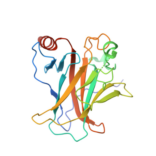
Structural Basis for Understanding Oncogenic P53 Mutations and Designing Rescue Drugs.

Joerger, A.C.Ang, H.C.Fersht, A.R.

(2006) Proc Natl Acad Sci U S A 103: 15056

  • DOI: https://doi.org/10.1073/pnas.0607286103
  • Primary Citation of Related Structures:  
    2J1W, 2J1X, 2J1Y, 2J1Z, 2J20, 2J21

  • PubMed Abstract: 

    The DNA-binding domain of the tumor suppressor p53 is inactivated by mutation in approximately 50% of human cancers. We have solved high-resolution crystal structures of several oncogenic mutants to investigate the structural basis of inactivation and provide information for designing drugs that may rescue inactivated mutants. We found a variety of structural consequences upon mutation: (i) the removal of an essential contact with DNA, (ii) creation of large, water-accessible crevices or hydrophobic internal cavities with no other structural changes but with a large loss of thermodynamic stability, (iii) distortion of the DNA-binding surface, and (iv) alterations to surfaces not directly involved in DNA binding but involved in domain-domain interactions on binding as a tetramer. These findings explain differences in functional properties and associated phenotypes (e.g., temperature sensitivity). Some mutants have the potential of being rescued by a generic stabilizing drug. In addition, a mutation-induced crevice is a potential target site for a mutant-selective stabilizing drug.


  • Organizational Affiliation

    Cambridge University Chemical Laboratory and Cambridge Centre for Protein Engineering, Medical Research Council Centre, Hills Road, Cambridge CB2 2QH, United Kingdom.


Macromolecules
Find similar proteins by:  (by identity cutoff)  |  3D Structure
Entity ID: 1
MoleculeChains Sequence LengthOrganismDetailsImage
CELLULAR TUMOR ANTIGEN P53
A, B, C, D
200Homo sapiensMutation(s): 5 
UniProt & NIH Common Fund Data Resources
Find proteins for P04637 (Homo sapiens)
Explore P04637 
Go to UniProtKB:  P04637
PHAROS:  P04637
GTEx:  ENSG00000141510 
Entity Groups  
Sequence Clusters30% Identity50% Identity70% Identity90% Identity95% Identity100% Identity
UniProt GroupP04637
Sequence Annotations
Expand
  • Reference Sequence
Experimental Data & Validation

Experimental Data

  • Method: X-RAY DIFFRACTION
  • Resolution: 1.69 Å
  • R-Value Free: 0.218 
  • R-Value Work: 0.189 
  • R-Value Observed: 0.189 
  • Space Group: P 1 21 1
Unit Cell:
Length ( Å )Angle ( ˚ )
a = 68.514α = 90
b = 73.076β = 90.05
c = 84.76γ = 90
Software Package:
Software NamePurpose
CNSrefinement
CNSphasing

Structure Validation

View Full Validation Report


Entry History 

Deposition Data

Revision History  (Full details and data files)

  • Version 1.0: 2006-09-20
    Type: Initial release
  • Version 1.1: 2011-05-08
    Changes: Version format compliance
  • Version 1.2: 2011-07-13
    Changes: Version format compliance
  • Version 1.3: 2023-12-13
    Changes: Data collection, Database references, Derived calculations, Other, Refinement description