2J0Q

The crystal structure of the Exon Junction Complex at 3.2 A resolution


Experimental Data Snapshot

  • Method: X-RAY DIFFRACTION
  • Resolution: 3.20 Å
  • R-Value Free: 0.277 
  • R-Value Work: 0.232 
  • R-Value Observed: 0.235 

wwPDB Validation   3D Report Full Report


Ligand Structure Quality Assessment 


This is version 1.2 of the entry. See complete history


Literature

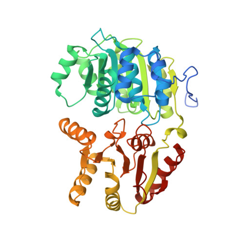
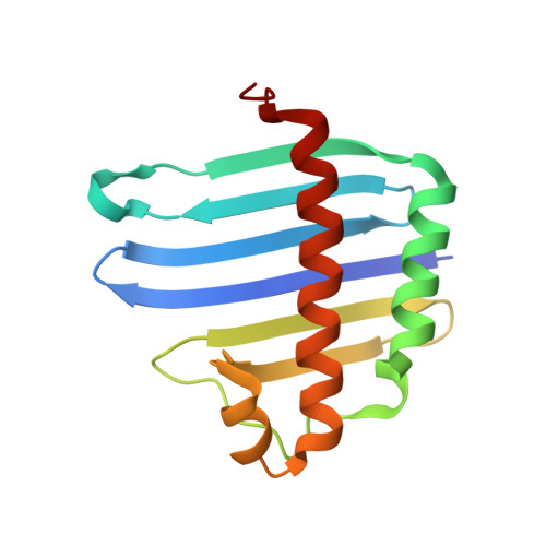
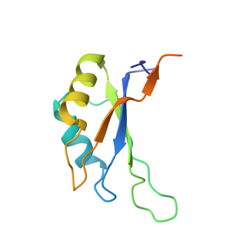
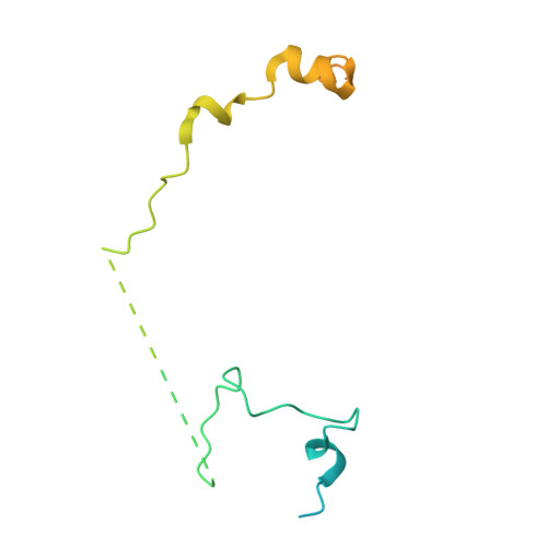
The Crystal Structure of the Exon Junction Complex Reveals How It Maintains a Stable Grip on Mrna.

Bono, F.Ebert, J.Lorentzen, E.Conti, E.

(2006) Cell 126: 713

  • DOI: https://doi.org/10.1016/j.cell.2006.08.006
  • Primary Citation of Related Structures:  
    2J0Q, 2J0S, 2J0U

  • PubMed Abstract: 

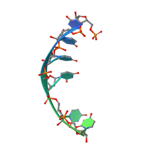
    The exon junction complex (EJC) plays a major role in posttranscriptional regulation of mRNA in metazoa. The EJC is deposited onto mRNA during splicing and is transported to the cytoplasm where it influences translation, surveillance, and localization of the spliced mRNA. The complex is formed by the association of four proteins (eIF4AIII, Barentsz [Btz], Mago, and Y14), mRNA, and ATP. The 2.2 A resolution structure of the EJC reveals how it stably locks onto mRNA. The DEAD-box protein eIF4AIII encloses an ATP molecule and provides the binding sites for six ribonucleotides. Btz wraps around eIF4AIII and stacks against the 5' nucleotide. An intertwined network of interactions anchors Mago-Y14 and Btz at the interface between the two domains of eIF4AIII, effectively stabilizing the ATP bound state. Comparison with the structure of the eIF4AIII-Btz subcomplex that we have also determined reveals that large conformational changes are required upon EJC assembly and disassembly.


  • Organizational Affiliation

    European Molecular Biology Laboratory, EMBL, Meyerhofstrasse 1, D-69117 Heidelberg, Germany.


Macromolecules

Find similar proteins by:  (by identity cutoff)  |  3D Structure
Entity ID: 1
MoleculeChains Sequence LengthOrganismDetailsImage
ATP-DEPENDENT RNA HELICASE DDX48
A, B
410Homo sapiensMutation(s): 0 
UniProt & NIH Common Fund Data Resources
Find proteins for P38919 (Homo sapiens)
Explore P38919 
Go to UniProtKB:  P38919
PHAROS:  P38919
GTEx:  ENSG00000141543 
Entity Groups  
Sequence Clusters30% Identity50% Identity70% Identity90% Identity95% Identity100% Identity
UniProt GroupP38919
Sequence Annotations
Expand
  • Reference Sequence
Find similar proteins by:  (by identity cutoff)  |  3D Structure
Entity ID: 2
MoleculeChains Sequence LengthOrganismDetailsImage
PROTEIN MAGO NASHI HOMOLOG
C, F
146Homo sapiensMutation(s): 0 
UniProt & NIH Common Fund Data Resources
Find proteins for P61326 (Homo sapiens)
Explore P61326 
Go to UniProtKB:  P61326
PHAROS:  P61326
GTEx:  ENSG00000162385 
Entity Groups  
Sequence Clusters30% Identity50% Identity70% Identity90% Identity95% Identity100% Identity
UniProt GroupP61326
Sequence Annotations
Expand
  • Reference Sequence
Find similar proteins by:  (by identity cutoff)  |  3D Structure
Entity ID: 3
MoleculeChains Sequence LengthOrganismDetailsImage
RNA-BINDING PROTEIN 8A
D, G
109Homo sapiensMutation(s): 0 
UniProt & NIH Common Fund Data Resources
Find proteins for Q9Y5S9 (Homo sapiens)
Explore Q9Y5S9 
Go to UniProtKB:  Q9Y5S9
PHAROS:  Q9Y5S9
GTEx:  ENSG00000265241 
Entity Groups  
Sequence Clusters30% Identity50% Identity70% Identity90% Identity95% Identity100% Identity
UniProt GroupQ9Y5S9
Sequence Annotations
Expand
  • Reference Sequence
Find similar proteins by:  (by identity cutoff)  |  3D Structure
Entity ID: 5
MoleculeChains Sequence LengthOrganismDetailsImage
PROTEIN CASC3I,
J [auth T]
150Homo sapiensMutation(s): 0 
UniProt & NIH Common Fund Data Resources
Find proteins for O15234 (Homo sapiens)
Explore O15234 
Go to UniProtKB:  O15234
PHAROS:  O15234
GTEx:  ENSG00000108349 
Entity Groups  
Sequence Clusters30% Identity50% Identity70% Identity90% Identity95% Identity100% Identity
UniProt GroupO15234
Sequence Annotations
Expand
  • Reference Sequence

Find similar nucleic acids by:  Sequence   |   3D Structure  

Entity ID: 4
MoleculeChains LengthOrganismImage
5'-R(*UP*UP*UP*UP*UP*UP*UP*UP*UP*UP*UP*UP*UP*UP*U)-3'
E, H
15N/A
Sequence Annotations
Expand
  • Reference Sequence
Experimental Data & Validation

Experimental Data

  • Method: X-RAY DIFFRACTION
  • Resolution: 3.20 Å
  • R-Value Free: 0.277 
  • R-Value Work: 0.232 
  • R-Value Observed: 0.235 
  • Space Group: P 21 21 21
Unit Cell:
Length ( Å )Angle ( ˚ )
a = 69.15α = 90
b = 161.24β = 90
c = 193.24γ = 90
Software Package:
Software NamePurpose
REFMACrefinement
XDSdata reduction
XSCALEdata scaling
PHASERphasing

Structure Validation

View Full Validation Report


Ligand Structure Quality Assessment 


Entry History 

Deposition Data

Revision History  (Full details and data files)

  • Version 1.0: 2006-08-30
    Type: Initial release
  • Version 1.1: 2011-07-13
    Changes: Advisory, Version format compliance
  • Version 1.2: 2023-12-13
    Changes: Data collection, Database references, Derived calculations, Other, Refinement description