2I5B

The crystal structure of an ADP complex of Bacillus subtilis pyridoxal kinase provides evidence for the parralel emergence of enzyme activity during evolution


Experimental Data Snapshot

  • Method: X-RAY DIFFRACTION
  • Resolution: 2.80 Å
  • R-Value Free: 0.274 
  • R-Value Work: 0.222 
  • R-Value Observed: 0.225 

wwPDB Validation   3D Report Full Report


Ligand Structure Quality Assessment 


This is version 1.4 of the entry. See complete history


Literature

The Crystal Structure of an ADP Complex of Bacillus subtilis Pyridoxal Kinase Provides Evidence for the Parallel Emergence of Enzyme Activity During Evolution.

Newman, J.A.Das, S.K.Sedelnikova, S.E.Rice, D.W.

(2006) J Mol Biol 363: 520-530

  • DOI: https://doi.org/10.1016/j.jmb.2006.08.013
  • Primary Citation of Related Structures:  
    2I5B

  • PubMed Abstract: 

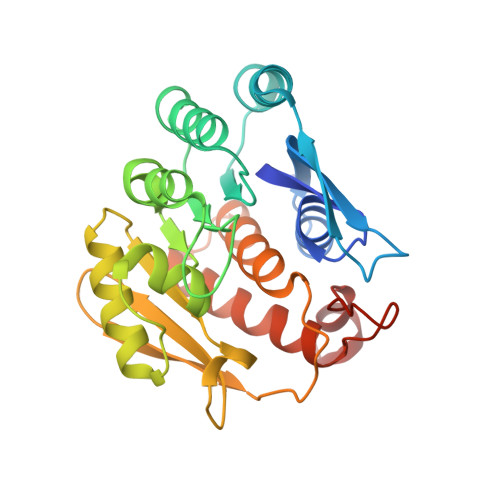
    Pyridoxal kinase catalyses the phosphorylation of pyridoxal, pyridoxine and pyridoxamine to their 5' phosphates and plays an important role in the pyridoxal 5' phosphate salvage pathway. The crystal structure of a dimeric pyridoxal kinase from Bacillus subtilis has been solved in complex with ADP to 2.8 A resolution. Analysis of the structure suggests that binding of the nucleotide induces the ordering of two loops, which operate independently to close a flap on the active site. Comparisons with other ribokinase superfamily members reveal that B. subtilis pyridoxal kinase is more closely related in both sequence and structure to the family of HMPP kinases than to other pyridoxal kinases, suggesting that this structure represents the first for a novel family of "HMPP kinase-like" pyridoxal kinases. Moreover this further suggests that this enzyme activity has evolved independently on multiple occasions from within the ribokinase superfamily.


  • Organizational Affiliation

    Krebs Institute for Biomolecular Research, Department of Molecular Biology and Biotechnology, The University of Sheffield, Sheffield S10 2TN, UK.


Macromolecules
Find similar proteins by:  (by identity cutoff)  |  3D Structure
Entity ID: 1
MoleculeChains Sequence LengthOrganismDetailsImage
Phosphomethylpyrimidine kinaseA,
B [auth C],
C [auth D],
D [auth B],
E
271Bacillus subtilisMutation(s): 0 
Gene Names: thiD
EC: 2.7.4.7
UniProt
Find proteins for P39610 (Bacillus subtilis (strain 168))
Explore P39610 
Go to UniProtKB:  P39610
Entity Groups  
Sequence Clusters30% Identity50% Identity70% Identity90% Identity95% Identity100% Identity
UniProt GroupP39610
Sequence Annotations
Expand
  • Reference Sequence
Experimental Data & Validation

Experimental Data

  • Method: X-RAY DIFFRACTION
  • Resolution: 2.80 Å
  • R-Value Free: 0.274 
  • R-Value Work: 0.222 
  • R-Value Observed: 0.225 
  • Space Group: P 41 21 2
Unit Cell:
Length ( Å )Angle ( ˚ )
a = 102.499α = 90
b = 102.499β = 90
c = 251.36γ = 90
Software Package:
Software NamePurpose
SCALAdata scaling
REFMACrefinement
PDB_EXTRACTdata extraction
MAR345data collection
MOSFLMdata reduction
CCP4data scaling
MOLREPphasing

Structure Validation

View Full Validation Report


Ligand Structure Quality Assessment 


Entry History 

Deposition Data

Revision History  (Full details and data files)

  • Version 1.0: 2006-09-19
    Type: Initial release
  • Version 1.1: 2008-05-01
    Changes: Version format compliance
  • Version 1.2: 2011-07-13
    Changes: Version format compliance
  • Version 1.3: 2017-10-18
    Changes: Refinement description
  • Version 1.4: 2023-08-30
    Changes: Data collection, Database references, Derived calculations, Refinement description