2HT6

Crystal structure of Human Gem G-domain bound to GDP


Experimental Data Snapshot

  • Method: X-RAY DIFFRACTION
  • Resolution: 2.40 Å
  • R-Value Free: 0.287 
  • R-Value Work: 0.226 
  • R-Value Observed: 0.229 

wwPDB Validation   3D Report Full Report


Ligand Structure Quality Assessment 


This is version 1.3 of the entry. See complete history


Literature

Structure-function studies of the G-domain from human gem, a novel small G-protein.

Opatowsky, Y.Sasson, Y.Shaked, I.Ward, Y.Chomsky-Hecht, O.Litvak, Y.Selinger, Z.Kelly, K.Hirsch, J.A.

(2006) FEBS Lett 580: 5959-5964

  • DOI: https://doi.org/10.1016/j.febslet.2006.09.067
  • Primary Citation of Related Structures:  
    2HT6

  • PubMed Abstract: 

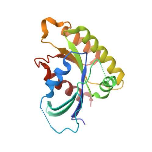
    Gem, a member of the Rad,Gem/Kir subfamily of small G-proteins, has unique sequence features. We report here the crystallographic structure determination of the Gem G-domain in complex with nucleotide to 2.4 A resolution. Although the basic Ras protein fold is maintained, the Gem switch regions emphatically differ from the Ras paradigm. Our ensuing biochemical characterization indicates that Gem G-domain markedly prefers GDP over GTP. Two known functions of Gem are distinctly affected by spatially separated clusters of mutations.


  • Organizational Affiliation

    Department of Biochemistry, Daniella Rich Institute for Structural Biology, Tel Aviv University, Ramat Aviv 69978, Israel.


Macromolecules
Find similar proteins by:  (by identity cutoff)  |  3D Structure
Entity ID: 1
MoleculeChains Sequence LengthOrganismDetailsImage
GTP-binding protein GEM
A, B
174Homo sapiensMutation(s): 0 
Gene Names: GEMKIR
UniProt & NIH Common Fund Data Resources
Find proteins for P55040 (Homo sapiens)
Explore P55040 
Go to UniProtKB:  P55040
PHAROS:  P55040
GTEx:  ENSG00000164949 
Entity Groups  
Sequence Clusters30% Identity50% Identity70% Identity90% Identity95% Identity100% Identity
UniProt GroupP55040
Sequence Annotations
Expand
  • Reference Sequence
Experimental Data & Validation

Experimental Data

  • Method: X-RAY DIFFRACTION
  • Resolution: 2.40 Å
  • R-Value Free: 0.287 
  • R-Value Work: 0.226 
  • R-Value Observed: 0.229 
  • Space Group: P 21 21 21
Unit Cell:
Length ( Å )Angle ( ˚ )
a = 39.843α = 90
b = 81.433β = 90
c = 124.095γ = 90
Software Package:
Software NamePurpose
REFMACrefinement
PDB_EXTRACTdata extraction
DENZOdata reduction
SCALEPACKdata scaling
SHARPphasing

Structure Validation

View Full Validation Report


Ligand Structure Quality Assessment 


Entry History 

Deposition Data

Revision History  (Full details and data files)

  • Version 1.0: 2006-10-31
    Type: Initial release
  • Version 1.1: 2008-05-01
    Changes: Version format compliance
  • Version 1.2: 2011-07-13
    Changes: Version format compliance
  • Version 1.3: 2024-02-14
    Changes: Data collection, Database references, Derived calculations, Refinement description