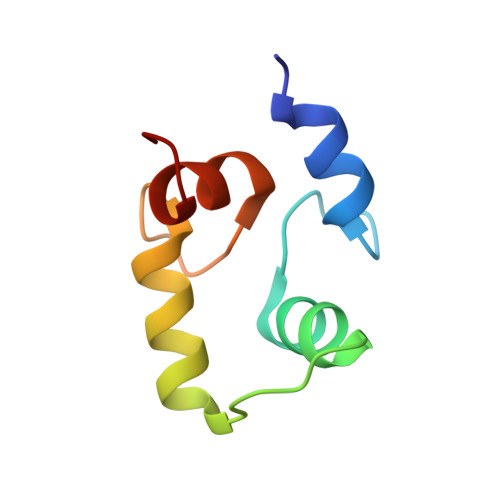
Calcium-induced folding of a fragment of calmodulin composed of EF-hands 2 and 3
Lakowski, T.M., Lee, G.M., Okon, M., Reid, R.E., McIntosh, L.P.(2007) Protein Sci 16: 1119-1132
- PubMed: 17473011
- DOI: https://doi.org/10.1110/ps.072777107
- Primary Citation of Related Structures:
2HF5 - PubMed Abstract:
Calmodulin (CaM) is an EF-hand protein composed of two calcium (Ca(2+))-binding EF-hand motifs in its N-domain (EF-1 and EF-2) and two in its C-domain (EF-3 and EF-4). In this study, we examined the structure, dynamics, and Ca(2+)-binding properties of a fragment of CaM containing only EF-2 and EF-3 and the intervening linker sequence (CaM2/3). Based on NMR spectroscopic analyses, Ca(2+)-free CaM2/3 is predominantly unfolded, but upon binding Ca(2+), adopts a monomeric structure composed of two EF-hand motifs bridged by a short antiparallel beta-sheet. Despite having an "even-odd" pairing of EF-hands, the tertiary structure of CaM2/3 is similar to both the "odd-even" paired N- and C-domains of Ca(2+)-ligated CaM, with the conformationally flexible linker sequence adopting the role of an inter-EF-hand loop. However, unlike either CaM domain, CaM2/3 exhibits stepwise Ca(2+) binding with a K (d1) = 30 +/- 5 microM to EF-3, and a K (d2) > 1000 microM to EF-2. Binding of the first equivalent of Ca(2+) induces the cooperative folding of CaM2/3. In the case of native CaM, stacking interactions between four conserved aromatic residues help to hold the first and fourth helices of each EF-hand domain together, while the loop between EF-hands covalently tethers the second and third helices. In contrast, these aromatic residues lie along the second and third helices of CaM2/3, and thus are positioned adjacent to the loop between its "even-odd" paired EF-hands. This nonnative hydrophobic core packing may contribute to the weak Ca(2+) affinity exhibited by EF-2 in the context of CaM2/3.
Organizational Affiliation:
Faculty of Pharmaceutical Sciences, Division of Biomolecular and Pharmaceutical Chemistry, University of British Columbia, Vancouver, British Columbia, Canada, V6T 1Z3.