2HD1

Crystal structure of PDE9 in complex with IBMX


Experimental Data Snapshot

  • Method: X-RAY DIFFRACTION
  • Resolution: 2.23 Å
  • R-Value Free: 0.231 
  • R-Value Work: 0.215 

wwPDB Validation   3D Report Full Report


Ligand Structure Quality Assessment 


This is version 1.4 of the entry. See complete history


Literature

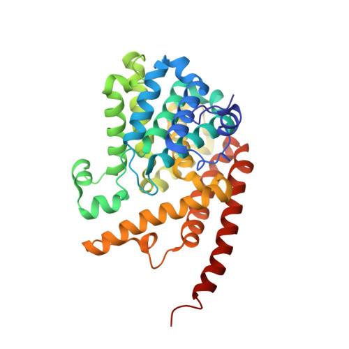
Crystal structure of phosphodiesterase 9 shows orientation variation of inhibitor IBMX binding

Huai, Q.Wang, H.Zhang, W.Colman, R.W.Robinson, H.Ke, H.

(2004) Proc Natl Acad Sci U S A 101: 9624-9629

  • DOI: https://doi.org/10.1073/pnas.0401120101
  • Primary Citation of Related Structures:  
    2HD1

  • PubMed Abstract: 

    Cyclic nucleotide phosphodiesterases (PDEs) are enzymes controlling cellular concentrations of the second messengers cAMP and cGMP. The crystal structure of the catalytic domain of PDE9A2, a member of a PDE family specifically hydrolyzing cGMP, has been determined at 2.23-A resolution. The PDE9A2 catalytic domain closely resembles the cAMP-specific PDE4D2 but is significantly different from the cGMP-specific PDE5A1, implying that each individual PDE family has its own characteristic substrate recognition mechanism. The different conformations of the H and M loops between PDE9A2 and PDE5A1 imply their less critical roles in nucleotide recognition. The nonselective inhibitor 3-isobutyl-1-methylxanthine (IBMX) binds to a similar subpocket in the active sites of PDE4, PDE5, and PDE9 and has a common pattern of the binding. However, significantly different orientations and interactions of IBMXs are observed among the three PDE families and also between two monomers of the PDE9A2 dimer. The kinetic properties of the PDE9A2 catalytic domain similar to those of full-length PDE9A imply that the N-terminal regulatory domain does not significantly alter the catalytic activity and the IBMX inhibition.


  • Organizational Affiliation

    Department of Biochemistry and Biophysics and Lineberger Comprehensive Cancer Center, University of North Carolina, Chapel Hill, NC 27599-7260, USA.


Macromolecules
Find similar proteins by:  (by identity cutoff)  |  3D Structure
Entity ID: 1
MoleculeChains Sequence LengthOrganismDetailsImage
Phosphodiesterase 9A
A, B
326Homo sapiensMutation(s): 0 
Gene Names: PDE
UniProt & NIH Common Fund Data Resources
Find proteins for O76083 (Homo sapiens)
Explore O76083 
Go to UniProtKB:  O76083
PHAROS:  O76083
GTEx:  ENSG00000160191 
Entity Groups  
Sequence Clusters30% Identity50% Identity70% Identity90% Identity95% Identity100% Identity
UniProt GroupO76083
Sequence Annotations
Expand
  • Reference Sequence
Small Molecules
Binding Affinity Annotations 
IDSourceBinding Affinity
IBM Binding MOAD:  2HD1 IC50: 5.00e+5 (nM) from 1 assay(s)
BindingDB:  2HD1 IC50: min: 1.17e+5, max: 2.00e+5 (nM) from 2 assay(s)
PDBBind:  2HD1 IC50: 5.00e+5 (nM) from 1 assay(s)
Experimental Data & Validation

Experimental Data

  • Method: X-RAY DIFFRACTION
  • Resolution: 2.23 Å
  • R-Value Free: 0.231 
  • R-Value Work: 0.215 
  • Space Group: P 41 21 2
Unit Cell:
Length ( Å )Angle ( ˚ )
a = 103.513α = 90
b = 103.513β = 90
c = 269.727γ = 90
Software Package:
Software NamePurpose
ADSCdata collection
DENZOdata reduction
AMoREphasing
CNSrefinement
SCALEPACKdata scaling

Structure Validation

View Full Validation Report


Ligand Structure Quality Assessment 


Entry History 

Deposition Data

Revision History  (Full details and data files)

  • Version 1.0: 2006-06-27
    Type: Initial release
  • Version 1.1: 2008-05-01
    Changes: Version format compliance
  • Version 1.2: 2011-07-13
    Changes: Version format compliance
  • Version 1.3: 2024-02-14
    Changes: Data collection, Database references, Derived calculations
  • Version 1.4: 2024-04-03
    Changes: Refinement description