2H59

Sir2 H116A-deacetylated p53 peptide-3'-o-acetyl ADP ribose


Experimental Data Snapshot

  • Method: X-RAY DIFFRACTION
  • Resolution: 1.90 Å
  • R-Value Free: 0.247 
  • R-Value Work: 0.205 
  • R-Value Observed: 0.207 

wwPDB Validation   3D Report Full Report


Ligand Structure Quality Assessment 


This is version 1.5 of the entry. See complete history


Literature

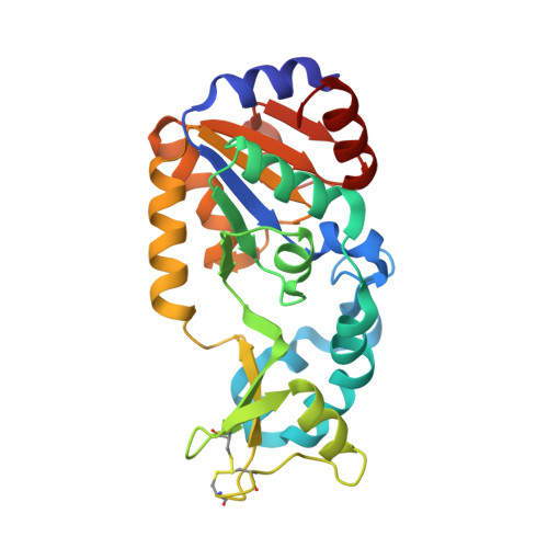
Insights into the Sirtuin Mechanism from Ternary Complexes Containing NAD(+) and Acetylated Peptide.

Hoff, K.G.Avalos, J.L.Sens, K.Wolberger, C.

(2006) Structure 14: 1231-1240

  • DOI: https://doi.org/10.1016/j.str.2006.06.006
  • Primary Citation of Related Structures:  
    2H4F, 2H4H, 2H4J, 2H59

  • PubMed Abstract: 

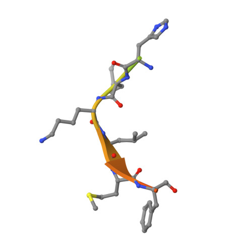
    Sirtuin proteins comprise a unique class of NAD+-dependent protein deacetylases. Although several structures of sirtuins have been determined, the mechanism by which NAD+ cleavage occurs has remained unclear. We report the structures of ternary complexes containing NAD+ and acetylated peptide bound to the bacterial sirtuin Sir2Tm and to a catalytic mutant (Sir2Tm(H116Y)). NAD+ in these structures binds in a conformation different from that seen in previous structures, exposing the alpha face of the nicotinamide ribose to the carbonyl oxygen of the acetyl lysine substrate. The NAD+ conformation is identical in both structures, suggesting that proper coenzyme orientation is not dependent on contacts with the catalytic histidine. We also present the structure of Sir2Tm(H116A) bound to deacteylated peptide and 3'-O-acetyl ADP ribose. Taken together, these structures suggest a mechanism for nicotinamide cleavage in which an invariant phenylalanine plays a central role in promoting formation of the O-alkylamidate reaction intermediate and preventing nicotinamide exchange.


  • Organizational Affiliation

    Department of Biophysics and Biophysical Chemistry, Howard Hughes Medical Institute, The Johns Hopkins University School of Medicine, 725 North Wolfe Street, Baltimore, Maryland 21205, USA.


Macromolecules
Find similar proteins by:  (by identity cutoff)  |  3D Structure
Entity ID: 1
MoleculeChains Sequence LengthOrganismDetailsImage
NAD-dependent deacetylase
A, B
246Thermotoga maritimaMutation(s): 1 
Gene Names: npdA
EC: 3.5.1
UniProt
Find proteins for Q9WYW0 (Thermotoga maritima (strain ATCC 43589 / DSM 3109 / JCM 10099 / NBRC 100826 / MSB8))
Explore Q9WYW0 
Go to UniProtKB:  Q9WYW0
Entity Groups  
Sequence Clusters30% Identity50% Identity70% Identity90% Identity95% Identity100% Identity
UniProt GroupQ9WYW0
Sequence Annotations
Expand
  • Reference Sequence

Find similar proteins by:  Sequence   |   3D Structure  

Entity ID: 2
MoleculeChains Sequence LengthOrganismDetailsImage
Cellular tumor antigen p53C [auth D],
D [auth E]
18N/AMutation(s): 0 
UniProt & NIH Common Fund Data Resources
Find proteins for P04637 (Homo sapiens)
Explore P04637 
Go to UniProtKB:  P04637
PHAROS:  P04637
GTEx:  ENSG00000141510 
Entity Groups  
Sequence Clusters30% Identity50% Identity70% Identity90% Identity95% Identity100% Identity
UniProt GroupP04637
Sequence Annotations
Expand
  • Reference Sequence
Small Molecules
Ligands 3 Unique
IDChains Name / Formula / InChI Key2D Diagram3D Interactions
3OD
Query on 3OD

Download Ideal Coordinates CCD File 
H [auth B](2S,3S,4R,5S)-2-({[(S)-{[(S)-{[(2R,3S,4R,5R)-5-(6-AMINO-9H-PURIN-9-YL)-3,4-DIHYDROXYTETRAHYDROFURAN-2-YL]METHOXY}(HYDROXY)PHOSPHORYL]OXY}(HYDROXY)PHOSPHORYL]OXY}METHYL)-4,5-DIHYDROXYTETRAHYDROFURAN-3-YL ACETATE
C17 H25 N5 O15 P2
HNHCIVXQBMBKPQ-ARMUENPQSA-N
APR
Query on APR

Download Ideal Coordinates CCD File 
F [auth A]ADENOSINE-5-DIPHOSPHORIBOSE
C15 H23 N5 O14 P2
SRNWOUGRCWSEMX-KEOHHSTQSA-N
ZN
Query on ZN

Download Ideal Coordinates CCD File 
E [auth A],
G [auth B]
ZINC ION
Zn
PTFCDOFLOPIGGS-UHFFFAOYSA-N
Experimental Data & Validation

Experimental Data

  • Method: X-RAY DIFFRACTION
  • Resolution: 1.90 Å
  • R-Value Free: 0.247 
  • R-Value Work: 0.205 
  • R-Value Observed: 0.207 
  • Space Group: P 41
Unit Cell:
Length ( Å )Angle ( ˚ )
a = 46.996α = 90
b = 46.996β = 90
c = 257.081γ = 90
Software Package:
Software NamePurpose
REFMACrefinement
Blu-Icedata collection
HKL-2000data scaling
MOLREPphasing

Structure Validation

View Full Validation Report


Ligand Structure Quality Assessment 


Entry History 

Deposition Data

Revision History  (Full details and data files)

  • Version 1.0: 2006-09-05
    Type: Initial release
  • Version 1.1: 2008-05-01
    Changes: Version format compliance
  • Version 1.2: 2011-07-13
    Changes: Version format compliance
  • Version 1.3: 2018-01-31
    Changes: Experimental preparation
  • Version 1.4: 2021-10-20
    Changes: Database references, Derived calculations
  • Version 1.5: 2023-08-30
    Changes: Data collection, Refinement description