2FJN

The structure of phosphotyrosine phosphatase 1B in complex with compound 2


Experimental Data Snapshot

  • Method: X-RAY DIFFRACTION
  • Resolution: 2.20 Å
  • R-Value Free: 0.244 
  • R-Value Work: 0.208 

wwPDB Validation   3D Report Full Report


Ligand Structure Quality Assessment 


This is version 1.4 of the entry. See complete history


Literature

Conformation-assisted inhibition of protein-tyrosine phosphatase-1B elicits inhibitor selectivity over T-cell protein-tyrosine phosphatase.

Asante-Appiah, E.Patel, S.Desponts, C.Taylor, J.M.Lau, C.Dufresne, C.Therien, M.Friesen, R.Becker, J.W.Leblanc, Y.Kennedy, B.P.Scapin, G.

(2006) J Biol Chem 281: 8010-8015

  • DOI: https://doi.org/10.1074/jbc.M511827200
  • Primary Citation of Related Structures:  
    2FJM, 2FJN

  • PubMed Abstract: 

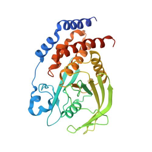
    PTP-1B represents an attractive target for the treatment of type 2 diabetes and obesity. Given the role that protein phosphatases play in the regulation of many biologically relevant processes, inhibitors against PTP-1B must be not only potent, but also selective. It has been extremely difficult to synthesize inhibitors that are selective over the highly homologous TCPTP. We have successfully exploited the conservative Leu119 to Val substitution between the two enzymes to synthesize a PTP-1B inhibitor that is an order of magnitude more selective over TCPTP. Structural analyses of PTP-1B/inhibitor complexes show a conformation-assisted inhibition mechanism as the basis for selectivity. Such an inhibitory mechanism may be applicable to other homologous enzymes.


  • Organizational Affiliation

    Department of Biochemistry and Molecular Biology, Merck Frosst Center for Therapeutic Research, Pointe-Claire, Dorval, Quebec H9R 4P8, Canada. ernest_asanteappiah@merck.com


Macromolecules
Find similar proteins by:  (by identity cutoff)  |  3D Structure
Entity ID: 1
MoleculeChains Sequence LengthOrganismDetailsImage
Tyrosine-protein phosphatase, non-receptor type 1
A, B
310Homo sapiensMutation(s): 0 
Gene Names: PTPN1PTP1B
EC: 3.1.3.48
UniProt & NIH Common Fund Data Resources
Find proteins for P18031 (Homo sapiens)
Explore P18031 
Go to UniProtKB:  P18031
PHAROS:  P18031
GTEx:  ENSG00000196396 
Entity Groups  
Sequence Clusters30% Identity50% Identity70% Identity90% Identity95% Identity100% Identity
UniProt GroupP18031
Sequence Annotations
Expand
  • Reference Sequence
Small Molecules
Ligands 2 Unique
IDChains Name / Formula / InChI Key2D Diagram3D Interactions
073
Query on 073

Download Ideal Coordinates CCD File 
D [auth A],
F [auth B]
(4-{(2S,4E)-2-(1H-1,2,3-BENZOTRIAZOL-1-YL)-2-[4-(METHOXYCARBONYL)PHENYL]-5-PHENYLPENT-4-ENYL}PHENYL)(DIFLUORO)METHYLPHOSPHONIC ACID
C32 H28 F2 N3 O5 P
GWWTUJWRRCTCSI-AOCUTKQBSA-N
CL
Query on CL

Download Ideal Coordinates CCD File 
C [auth A],
E [auth B]
CHLORIDE ION
Cl
VEXZGXHMUGYJMC-UHFFFAOYSA-M
Binding Affinity Annotations 
IDSourceBinding Affinity
073 BindingDB:  2FJN IC50: min: 29, max: 142 (nM) from 6 assay(s)
Binding MOAD:  2FJN IC50: 39 (nM) from 1 assay(s)
Experimental Data & Validation

Experimental Data

  • Method: X-RAY DIFFRACTION
  • Resolution: 2.20 Å
  • R-Value Free: 0.244 
  • R-Value Work: 0.208 
  • Space Group: P 21 21 21
Unit Cell:
Length ( Å )Angle ( ˚ )
a = 87.429α = 90
b = 85.617β = 90
c = 137.272γ = 90
Software Package:
Software NamePurpose
X-GENdata scaling
X-GENdata reduction
CNXrefinement
CNXphasing

Structure Validation

View Full Validation Report


Ligand Structure Quality Assessment 


Entry History 

Revision History  (Full details and data files)

  • Version 1.0: 2006-01-17
    Type: Initial release
  • Version 1.1: 2008-05-01
    Changes: Version format compliance
  • Version 1.2: 2011-07-13
    Changes: Version format compliance
  • Version 1.3: 2017-10-18
    Changes: Refinement description
  • Version 1.4: 2023-08-30
    Changes: Data collection, Database references, Derived calculations, Refinement description