2F3Y

Calmodulin/IQ domain complex


Experimental Data Snapshot

  • Method: X-RAY DIFFRACTION
  • Resolution: 1.45 Å
  • R-Value Free: 0.219 
  • R-Value Work: 0.189 
  • R-Value Observed: 0.189 

wwPDB Validation   3D Report Full Report


This is version 1.4 of the entry. See complete history


Literature

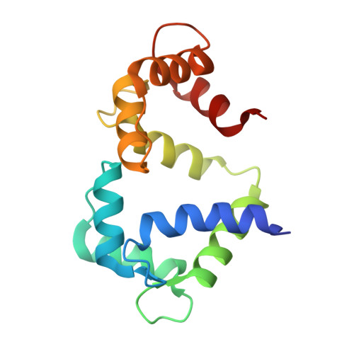
Structure of Calmodulin Bound to the Hydrophobic IQ Domain of the Cardiac Ca(v)1.2 Calcium Channel.

Fallon, J.L.Halling, D.B.Hamilton, S.L.Quiocho, F.A.

(2005) Structure 13: 1881-1886

  • DOI: https://doi.org/10.1016/j.str.2005.09.021
  • Primary Citation of Related Structures:  
    2F3Y, 2F3Z

  • PubMed Abstract: 

    Ca2+-dependent inactivation (CDI) and facilitation (CDF) of the Ca(v)1.2 Ca2+ channel require calmodulin binding to a putative IQ motif in the carboxy-terminal tail of the pore-forming subunit. We present the 1.45 A crystal structure of Ca2+-calmodulin bound to a 21 residue peptide corresponding to the IQ domain of Ca(v)1.2. This structure shows that parallel binding of calmodulin to the IQ domain is governed by hydrophobic interactions. Mutations of residues I1672 and Q1673 in the peptide to alanines, which abolish CDI but not CDF in the channel, do not greatly alter the structure. Both lobes of Ca2+-saturated CaM bind to the IQ peptide but isoleucine 1672, thought to form an intramolecular interaction that drives CDI, is buried. These findings suggest that this structure could represent the conformation that calmodulin assumes in CDF.


  • Organizational Affiliation

    Verna and Marrs McLean Department of Biochemistry and Molecular Biology, Baylor College of Medicine, Houston, Texas 77030, USA.


Macromolecules
Find similar proteins by:  (by identity cutoff)  |  3D Structure
Entity ID: 1
MoleculeChains Sequence LengthOrganismDetailsImage
Calmodulin148Homo sapiensMutation(s): 0 
Gene Names: calm1calm2calm3
UniProt & NIH Common Fund Data Resources
Find proteins for P0DP23 (Homo sapiens)
Explore P0DP23 
Go to UniProtKB:  P0DP23
PHAROS:  P0DP23
GTEx:  ENSG00000198668 
Entity Groups  
Sequence Clusters30% Identity50% Identity70% Identity90% Identity95% Identity100% Identity
UniProt GroupP0DP23
Sequence Annotations
Expand
  • Reference Sequence

Find similar proteins by:  Sequence   |   3D Structure  

Entity ID: 2
MoleculeChains Sequence LengthOrganismDetailsImage
Voltage-dependent L-type calcium channel alpha-1C subunit21N/AMutation(s): 0 
UniProt & NIH Common Fund Data Resources
Find proteins for Q13936 (Homo sapiens)
Explore Q13936 
Go to UniProtKB:  Q13936
PHAROS:  Q13936
GTEx:  ENSG00000151067 
Entity Groups  
Sequence Clusters30% Identity50% Identity70% Identity90% Identity95% Identity100% Identity
UniProt GroupQ13936
Sequence Annotations
Expand
  • Reference Sequence
Experimental Data & Validation

Experimental Data

  • Method: X-RAY DIFFRACTION
  • Resolution: 1.45 Å
  • R-Value Free: 0.219 
  • R-Value Work: 0.189 
  • R-Value Observed: 0.189 
  • Space Group: C 1 2 1
Unit Cell:
Length ( Å )Angle ( ˚ )
a = 86.2α = 90
b = 30.96β = 114.54
c = 63.94γ = 90
Software Package:
Software NamePurpose
DENZOdata reduction
SCALEPACKdata scaling
CNSphasing
CNSrefinement
PDB_EXTRACTdata extraction
MAR345data collection

Structure Validation

View Full Validation Report


Entry History 

Deposition Data

Revision History  (Full details and data files)

  • Version 1.0: 2005-12-27
    Type: Initial release
  • Version 1.1: 2008-05-01
    Changes: Version format compliance
  • Version 1.2: 2011-07-13
    Changes: Version format compliance
  • Version 1.3: 2017-10-18
    Changes: Refinement description
  • Version 1.4: 2024-02-14
    Changes: Data collection, Database references, Derived calculations