2EUF

X-ray structure of human CDK6-Vcyclin in complex with the inhibitor PD0332991


Experimental Data Snapshot

  • Method: X-RAY DIFFRACTION
  • Resolution: 3.00 Å
  • R-Value Free: 0.306 
  • R-Value Work: 0.229 
  • R-Value Observed: 0.233 

wwPDB Validation   3D Report Full Report


Ligand Structure Quality Assessment 


This is version 1.3 of the entry. See complete history


Literature

Toward understanding the structural basis of cyclin-dependent kinase 6 specific inhibition.

Lu, H.Schulze-Gahmen, U.

(2006) J Med Chem 49: 3826-3831

  • DOI: https://doi.org/10.1021/jm0600388
  • Primary Citation of Related Structures:  
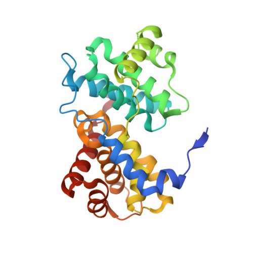
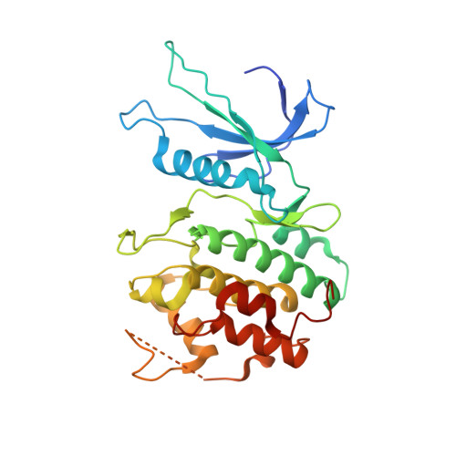
    2EUF, 2F2C

  • PubMed Abstract: 

    Cyclin-dependent kinases (CDKs) are key players in cell cycle control, and genetic alterations of CDKs and their regulators have been linked to a variety of cancers. Hence, CDKs are obvious targets for therapeutic intervention in various proliferative diseases, including cancer. To date, drug design efforts have mostly focused on CDK2 because methods for crystallization of its inhibitor complexes have been well established. CDK4 and CDK6, however, may be at least as important as enzymes for cell cycle regulation and could provide alternative treatment options. We describe here two complex structures of human CDK6 with a very specific kinase inhibitor, PD0332991, which is based on a pyrido[2,3-d]pyrimidin-7-one scaffold, and with the less specific aminopurvalanol inhibitor. Analysis of the structures suggests that relatively small conformational differences between CDK2 and CDK6 in the hinge region are contributing to the inhibitor specificity by inducing changes in the inhibitor orientation that lead to sterical clashes in CDK2 but not CDK6. These complex structures provide valuable insights for the future development of CDK-specific inhibitors.


  • Organizational Affiliation

    Physical Biosciences Division at Lawrence Berkeley National Laboratory, 1 Cyclotron Road, MS3, Berkeley, California, USA.


Macromolecules
Find similar proteins by:  (by identity cutoff)  |  3D Structure
Entity ID: 1
MoleculeChains Sequence LengthOrganismDetailsImage
viral Cyclin254Herpesvirus saimiri (strain 11)Mutation(s): 0 
Gene Names: 72ECLF2
UniProt
Find proteins for Q01043 (Saimiriine herpesvirus 2 (strain 11))
Explore Q01043 
Go to UniProtKB:  Q01043
Entity Groups  
Sequence Clusters30% Identity50% Identity70% Identity90% Identity95% Identity100% Identity
UniProt GroupQ01043
Sequence Annotations
Expand
  • Reference Sequence
Find similar proteins by:  (by identity cutoff)  |  3D Structure
Entity ID: 2
MoleculeChains Sequence LengthOrganismDetailsImage
Cell division protein kinase 6308Homo sapiensMutation(s): 0 
Gene Names: CDK6
EC: 2.7.1.37
UniProt & NIH Common Fund Data Resources
Find proteins for Q00534 (Homo sapiens)
Explore Q00534 
Go to UniProtKB:  Q00534
PHAROS:  Q00534
GTEx:  ENSG00000105810 
Entity Groups  
Sequence Clusters30% Identity50% Identity70% Identity90% Identity95% Identity100% Identity
UniProt GroupQ00534
Sequence Annotations
Expand
  • Reference Sequence
Small Molecules
Ligands 4 Unique
IDChains Name / Formula / InChI Key2D Diagram3D Interactions
LQQ
Query on LQQ

Download Ideal Coordinates CCD File 
E [auth B]6-ACETYL-8-CYCLOPENTYL-5-METHYL-2-[(5-PIPERAZIN-1-YLPYRIDIN-2-YL)AMINO]PYRIDO[2,3-D]PYRIMIDIN-7(8H)-ONE
C24 H29 N7 O2
AHJRHEGDXFFMBM-UHFFFAOYSA-N
DMS
Query on DMS

Download Ideal Coordinates CCD File 
F [auth B]DIMETHYL SULFOXIDE
C2 H6 O S
IAZDPXIOMUYVGZ-UHFFFAOYSA-N
ACT
Query on ACT

Download Ideal Coordinates CCD File 
D [auth B]ACETATE ION
C2 H3 O2
QTBSBXVTEAMEQO-UHFFFAOYSA-M
CA
Query on CA

Download Ideal Coordinates CCD File 
C [auth A]CALCIUM ION
Ca
BHPQYMZQTOCNFJ-UHFFFAOYSA-N
Binding Affinity Annotations 
IDSourceBinding Affinity
LQQ BindingDB:  2EUF IC50: min: 15, max: 21 (nM) from 4 assay(s)
PDBBind:  2EUF IC50: 15 (nM) from 1 assay(s)
Experimental Data & Validation

Experimental Data

  • Method: X-RAY DIFFRACTION
  • Resolution: 3.00 Å
  • R-Value Free: 0.306 
  • R-Value Work: 0.229 
  • R-Value Observed: 0.233 
  • Space Group: P 65 2 2
Unit Cell:
Length ( Å )Angle ( ˚ )
a = 71.146α = 90
b = 71.146β = 90
c = 446.876γ = 120
Software Package:
Software NamePurpose
REFMACrefinement
HKL-2000data reduction
CCP4data scaling
AMoREphasing

Structure Validation

View Full Validation Report


Ligand Structure Quality Assessment 


Entry History 

Deposition Data

Revision History  (Full details and data files)

  • Version 1.0: 2006-07-04
    Type: Initial release
  • Version 1.1: 2008-05-01
    Changes: Version format compliance
  • Version 1.2: 2011-07-13
    Changes: Advisory, Version format compliance
  • Version 1.3: 2024-02-14
    Changes: Data collection, Database references, Derived calculations