


Structures of Two Human Ubiquitin-Conjugating Enzymes from Twinned Crystals
Dodd, R.B., Read, R.J.To be published.
Experimental Data Snapshot
wwPDB Validation 3D Report Full Report

Entity ID: 1 | |||||
|---|---|---|---|---|---|
| Molecule | Chains | Sequence Length | Organism | Details | Image |
| UBIQUITIN-CONJUGATING ENZYME E2 D1 | 165 | Homo sapiens | Mutation(s): 0 EC: 6.3.2.19 | 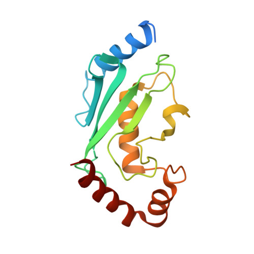 | |
UniProt & NIH Common Fund Data Resources | |||||
Find proteins for P51668 (Homo sapiens) Explore P51668 Go to UniProtKB: P51668 | |||||
PHAROS: P51668 GTEx: ENSG00000072401 | |||||
Entity Groups | |||||
| Sequence Clusters | 30% Identity50% Identity70% Identity90% Identity95% Identity100% Identity | ||||
| UniProt Group | P51668 | ||||
Sequence AnnotationsExpand | |||||
| |||||
| Length ( Å ) | Angle ( ˚ ) |
|---|---|
| a = 45.621 | α = 90 |
| b = 50.565 | β = 90 |
| c = 66.043 | γ = 90 |
| Software Name | Purpose |
|---|---|
| CNS | refinement |
| MOSFLM | data reduction |
| SCALA | data scaling |
| PHASER | phasing |
RCSB PDB (citation) is hosted by
RCSB PDB is a member of the

