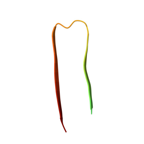
3D structure of Alzheimer's amyloid-{beta}(1-42) fibrils.
Luhrs, T., Ritter, C., Adrian, M., Riek-Loher, D., Bohrmann, B., Dobeli, H., Schubert, D., Riek, R.(2005) Proc Natl Acad Sci U S A 102: 17342-17347
- PubMed: 16293696
- DOI: https://doi.org/10.1073/pnas.0506723102
- Primary Citation of Related Structures:
2BEG - PubMed Abstract:
Alzheimer's disease is the most fatal neurodegenerative disorder wherein the process of amyloid-beta (Abeta) amyloidogenesis appears causative. Here, we present the 3D structure of the fibrils comprising Abeta(1-42), which was obtained by using hydrogen-bonding constraints from quenched hydrogen/deuterium-exchange NMR, side-chain packing constraints from pairwise mutagenesis studies, and parallel, in-register beta-sheet arrangement from previous solid-state NMR studies. Although residues 1-17 are disordered, residues 18-42 form a beta-strand-turn-beta-strand motif that contains two intermolecular, parallel, in-register beta-sheets that are formed by residues 18-26 (beta1) and 31-42 (beta2). At least two molecules of Abeta(1-42) are required to achieve the repeating structure of a protofilament. Intermolecular side-chain contacts are formed between the odd-numbered residues of strand beta1 of the nth molecule and the even-numbered residues of strand beta2 of the (n - 1)th molecule. This interaction pattern leads to partially unpaired beta-strands at the fibrillar ends, which explains the sequence selectivity, the cooperativity, and the apparent unidirectionality of Abeta fibril growth. It also provides a structural basis for fibrillization inhibitors.
Organizational Affiliation:
The Salk Institute for Biological Studies, La Jolla, CA 92037, USA.


