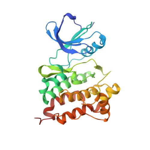
Structural basis of Src tyrosine kinase inhibition with a new class of potent and selective trisubstituted purine-based compounds.
Dalgarno, D., Stehle, T., Narula, S., Schelling, P., van Schravendijk, M.R., Adams, S., Andrade, L., Keats, J., Ram, M., Jin, L., Grossman, T., MacNeil, I., Metcalf III, C., Shakespeare, W., Wang, Y., Keenan, T., Sundaramoorthi, R., Bohacek, R., Weigele, M., Sawyer, T.(2006) Chem Biol Drug Des 67: 46-57
- PubMed: 16492148
- DOI: https://doi.org/10.1111/j.1747-0285.2005.00316.x
- Primary Citation of Related Structures:
2BDF, 2BDJ - PubMed Abstract:
The tyrosine kinase pp60src (Src) is the prototypical member of a family of proteins that participate in a broad array of cellular signal transduction processes, including cell growth, differentiation, survival, adhesion, and migration. Abnormal Src family kinase (SFK) signaling has been linked to several disease states, including osteoporosis and cancer metastases. Src has thus emerged as a molecular target for the discovery of small-molecule inhibitors that regulate Src kinase activity by binding to the ATP pocket within the catalytic domain. Here, we present crystal structures of the kinase domain of Src in complex with two purine-based inhibitors: AP23451, a small-molecule inhibitor designed to inhibit Src-dependent bone resorption, and AP23464, a small-molecule inhibitor designed to inhibit the Src-dependent metastatic spread of cancer. In each case, a trisubstituted purine template core was elaborated using structure-based drug design to yield a potent Src kinase inhibitor. These structures represent early examples of high affinity purine-based Src family kinase-inhibitor complexes, and they provide a detailed view of the specific protein-ligand interactions that lead to potent inhibition of Src. In particular, the 3-hydroxyphenethyl N9 substituent of AP23464 forms unique interactions with the protein that are critical to the picomolar affinity of this compound for Src. The comparison of these new structures with two relevant kinase-inhibitor complexes provides a structural basis for the observed kinase inhibitory selectivity. Further comparisons reveal a concerted induced-fit movement between the N- and C-terminal lobes of the kinase that correlates with the affinity of the ligand. Binding of the most potent inhibitor, AP23464, results in the largest induced-fit movement, which can be directly linked to interactions of the hydrophenethyl N9 substituent with a region at the interface between the two lobes. A less pronounced induced-fit movement is also observed in the Src-AP23451 complex. These new structures illustrate how the combination of structural, computational, and medicinal chemistry can be used to rationalize the process of developing high affinity, selective tyrosine kinase inhibitors as potential therapeutic agents.
Organizational Affiliation:
ARIAD Pharmaceuticals, 26 Landsdowne Street, Cambridge, MA 02139, USA. dalgarno@ariad.com