2BAO

Solution NMR structure of the myristoylated N-terminal fragment of Arf6


Experimental Data Snapshot

  • Method: SOLUTION NMR
  • Conformers Calculated: 50 
  • Conformers Submitted: 12 
  • Selection Criteria: structures with the lowest energy 

wwPDB Validation   3D Report Full Report


This is version 1.3 of the entry. See complete history


Literature

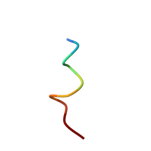
NMR structural studies of the myristoylated N-terminus of ADP ribosylation factor 6 (Arf6).

Gizachew, D.Oswald, R.

(2006) FEBS Lett 580: 4296-4301

  • DOI: https://doi.org/10.1016/j.febslet.2006.06.086
  • Primary Citation of Related Structures:  
    2BAO, 2BAU

  • PubMed Abstract: 

    Arf proteins are guanine nucleotide binding proteins that are implicated in endocytotic pathways and vesicle trafficking. The two widely studied isoforms of Arf proteins (Arf1 and Arf6) have different cellular functions and localizations but similar structures. Arf proteins have an N-terminal helix with a covalently bound myristoyl group. Except structural models, there are no three dimensional structures of the myristoylated N-terminal peptide or the intact myristoylated Arf proteins. However, understanding the role of both the myristoyl group and the N-terminal helix based on the details of their molecular structures is of great interest. In the solution structure of myristoylated N-terminal peptide of Arf6 described here, the myristoyl group folds toward the N-terminus to interact with the hydrophobic residues in particular, the phenyl ring. Also, the structure of the dodecylphosphocholine (DPC) micelle-bound of the peptide together with paramagnetic studies showed that the myristoyl group is inserted into the micelle while residues V4-G10 interact with the surface of the micelle. The structural differences between the unbound and micelle-bound myristoylated N-terminal peptide of Arf6 involves the myristoyl group and the side chains of the hydrophobic residues.


  • Organizational Affiliation

    Department of Molecular Medicine, Cornell University, Ithaca, NY 14853, USA. dg58@cornell.edu


Macromolecules

Find similar proteins by:  Sequence   |   3D Structure  

Entity ID: 1
MoleculeChains Sequence LengthOrganismDetailsImage
ADP-ribosylation factor 610N/AMutation(s): 0 
Membrane Entity: Yes 
UniProt & NIH Common Fund Data Resources
Find proteins for P62330 (Homo sapiens)
Explore P62330 
Go to UniProtKB:  P62330
PHAROS:  P62330
GTEx:  ENSG00000165527 
Entity Groups  
Sequence Clusters30% Identity50% Identity70% Identity90% Identity95% Identity100% Identity
UniProt GroupP62330
Sequence Annotations
Expand
  • Reference Sequence
Small Molecules
Ligands 1 Unique
IDChains Name / Formula / InChI Key2D Diagram3D Interactions
MYR
Query on MYR

Download Ideal Coordinates CCD File 
B [auth A]MYRISTIC ACID
C14 H28 O2
TUNFSRHWOTWDNC-UHFFFAOYSA-N
Experimental Data & Validation

Experimental Data

  • Method: SOLUTION NMR
  • Conformers Calculated: 50 
  • Conformers Submitted: 12 
  • Selection Criteria: structures with the lowest energy 

Structure Validation

View Full Validation Report


Entry History 

Deposition Data

  • Released Date: 2006-07-25 
  • Deposition Author(s): Gizachew, D.

Revision History  (Full details and data files)

  • Version 1.0: 2006-07-25
    Type: Initial release
  • Version 1.1: 2008-05-01
    Changes: Version format compliance
  • Version 1.2: 2011-07-13
    Changes: Version format compliance
  • Version 1.3: 2022-03-09
    Changes: Database references, Derived calculations