2A8J

Crystal Structure of human Taspase1 (acivated form)


Experimental Data Snapshot

  • Method: X-RAY DIFFRACTION
  • Resolution: 1.90 Å
  • R-Value Free: 0.269 
  • R-Value Work: 0.223 
  • R-Value Observed: 0.223 

wwPDB Validation   3D Report Full Report


This is version 1.3 of the entry. See complete history


Literature

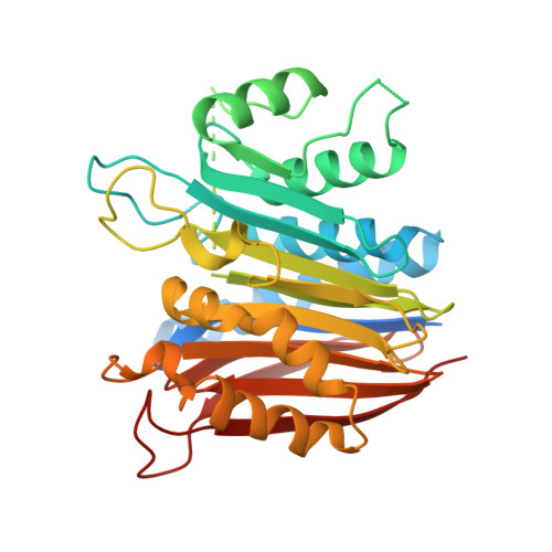
Crystal Structure of Human Taspase1, a Crucial Protease Regulating the Function of MLL.

Khan, J.A.Dunn, B.M.Tong, L.

(2005) Structure 13: 1443-1452

  • DOI: https://doi.org/10.1016/j.str.2005.07.006
  • Primary Citation of Related Structures:  
    2A8I, 2A8J, 2A8L, 2A8M

  • PubMed Abstract: 

    Taspase1 catalyzes the proteolytic processing of the mixed lineage leukemia (MLL) nuclear protein, which is required for maintaining Hox gene expression patterns. Chromosomal translocations of the MLL gene are associated with leukemia in infants. Taspase1, a threonine aspartase, is a member of the type 2 asparaginase family, but is the only protease in this family. We report here the crystal structures of human activated Taspase1 and its proenzyme, as well as the characterization of the effects of mutations in the active site region using a newly developed fluorogenic assay. The structure of Taspase1 has significant differences from other asparaginases, especially near the active site. Mutation of the catalytic nucleophile, Thr234, abolishes autocatalytic processing in cis but does not completely block proteolysis in trans. The structure unexpectedly showed the binding of a chloride ion in the active site, and our kinetic studies confirm that chlorides ions are inhibitors of this enzyme at physiologically relevant concentrations.


  • Organizational Affiliation

    Department of Biological Sciences, Columbia University, New York, New York 10027, USA.


Macromolecules
Find similar proteins by:  (by identity cutoff)  |  3D Structure
Entity ID: 1
MoleculeChains Sequence LengthOrganismDetailsImage
Threonine aspartase 1
A, B
420Homo sapiensMutation(s): 0 
Gene Names: C20orf13
EC: 3.4.25
UniProt & NIH Common Fund Data Resources
Find proteins for Q9H6P5 (Homo sapiens)
Explore Q9H6P5 
Go to UniProtKB:  Q9H6P5
PHAROS:  Q9H6P5
GTEx:  ENSG00000089123 
Entity Groups  
Sequence Clusters30% Identity50% Identity70% Identity90% Identity95% Identity100% Identity
UniProt GroupQ9H6P5
Sequence Annotations
Expand
  • Reference Sequence
Small Molecules
Ligands 1 Unique
IDChains Name / Formula / InChI Key2D Diagram3D Interactions
CL
Query on CL

Download Ideal Coordinates CCD File 
C [auth A],
D [auth B]
CHLORIDE ION
Cl
VEXZGXHMUGYJMC-UHFFFAOYSA-M
Experimental Data & Validation

Experimental Data

  • Method: X-RAY DIFFRACTION
  • Resolution: 1.90 Å
  • R-Value Free: 0.269 
  • R-Value Work: 0.223 
  • R-Value Observed: 0.223 
  • Space Group: P 21 21 21
Unit Cell:
Length ( Å )Angle ( ˚ )
a = 60.574α = 90
b = 89.659β = 90
c = 104.955γ = 90
Software Package:
Software NamePurpose
CNSrefinement
DENZOdata reduction
SCALEPACKdata scaling
COMOphasing

Structure Validation

View Full Validation Report


Entry History 

Deposition Data

Revision History  (Full details and data files)

  • Version 1.0: 2005-10-25
    Type: Initial release
  • Version 1.1: 2008-04-30
    Changes: Version format compliance
  • Version 1.2: 2011-07-13
    Changes: Version format compliance
  • Version 1.3: 2024-02-14
    Changes: Data collection, Database references, Derived calculations