1FCJ

CRYSTAL STRUCTURE OF OASS COMPLEXED WITH CHLORIDE AND SULFATE


Experimental Data Snapshot

  • Method: X-RAY DIFFRACTION
  • Resolution: 2.00 Å
  • R-Value Free: 0.246 
  • R-Value Work: 0.200 
  • R-Value Observed: 0.200 

wwPDB Validation   3D Report Full Report


Ligand Structure Quality Assessment 


This is version 1.3 of the entry. See complete history


Literature

Identification of an allosteric anion-binding site on O-acetylserine sulfhydrylase: structure of the enzyme with chloride bound.

Burkhard, P.Tai, C.H.Jansonius, J.N.Cook, P.F.

(2000) J Mol Biol 303: 279-286

  • DOI: https://doi.org/10.1006/jmbi.2000.4109
  • Primary Citation of Related Structures:  
    1FCJ

  • PubMed Abstract: 

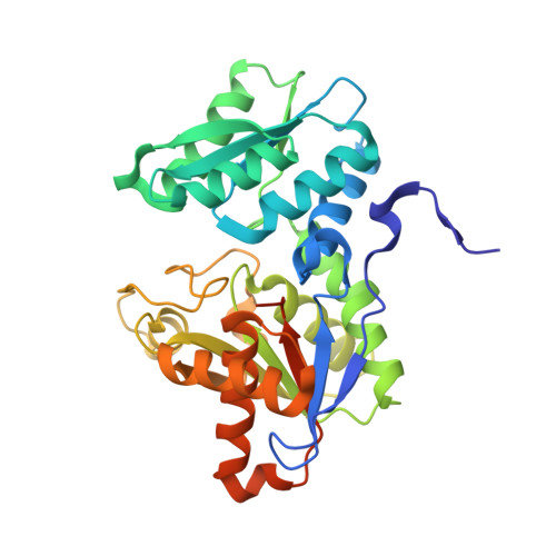
    A new crystal structure of O-acetylserine sulfhydrylase (OASS) has been solved with chloride bound at an allosteric site and sulfate bound at the active site. The bound anions result in a new "inhibited" conformation, that differs from the "open" native or "closed" external aldimine conformations. The allosteric site is located at the OASS dimer interface. The new inhibited structure involves a change in the position of the "moveable domain" (residues 87-131) to a location that differs from that in the open or closed forms. Formation of the external aldimine with substrate is stabilized by interaction of the alpha-carboxyl group of the substrate with a substrate-binding loop that is part of the moveable domain. The inhibited conformation prevents the substrate-binding loop from interacting with the alpha-carboxyl group, and hinders formation of the external Schiff base and thus subsequent chemistry. Chloride may be an analog of sulfide, the physiological inhibitor. Finally, these results suggest that OASS represents a new class of PLP-dependent enzymes that is regulated by small anions.


  • Organizational Affiliation

    M.E. Müller Institute for Structural Biology. Peter.Burkhard@unibas.ch


Macromolecules
Find similar proteins by:  (by identity cutoff)  |  3D Structure
Entity ID: 1
MoleculeChains Sequence LengthOrganismDetailsImage
O-ACETYLSERINE SULFHYDRYLASE
A, B, C, D
322Salmonella enterica subsp. enterica serovar TyphimuriumMutation(s): 0 
EC: 4.2.99.8
UniProt
Find proteins for P0A1E3 (Salmonella typhimurium (strain LT2 / SGSC1412 / ATCC 700720))
Explore P0A1E3 
Go to UniProtKB:  P0A1E3
Entity Groups  
Sequence Clusters30% Identity50% Identity70% Identity90% Identity95% Identity100% Identity
UniProt GroupP0A1E3
Sequence Annotations
Expand
  • Reference Sequence
Small Molecules
Ligands 3 Unique
IDChains Name / Formula / InChI Key2D Diagram3D Interactions
PLP
Query on PLP

Download Ideal Coordinates CCD File 
G [auth A],
J [auth B],
M [auth C],
P [auth D]
PYRIDOXAL-5'-PHOSPHATE
C8 H10 N O6 P
NGVDGCNFYWLIFO-UHFFFAOYSA-N
SO4
Query on SO4

Download Ideal Coordinates CCD File 
E [auth A],
H [auth B],
K [auth C],
N [auth D]
SULFATE ION
O4 S
QAOWNCQODCNURD-UHFFFAOYSA-L
CL
Query on CL

Download Ideal Coordinates CCD File 
F [auth A],
I [auth B],
L [auth C],
O [auth D]
CHLORIDE ION
Cl
VEXZGXHMUGYJMC-UHFFFAOYSA-M
Experimental Data & Validation

Experimental Data

  • Method: X-RAY DIFFRACTION
  • Resolution: 2.00 Å
  • R-Value Free: 0.246 
  • R-Value Work: 0.200 
  • R-Value Observed: 0.200 
  • Space Group: P 21 21 21
Unit Cell:
Length ( Å )Angle ( ˚ )
a = 96.38α = 90
b = 100.49β = 90
c = 142.81γ = 90
Software Package:
Software NamePurpose
AMoREphasing
CNSrefinement
MOSFLMdata reduction
CCP4data scaling

Structure Validation

View Full Validation Report


Ligand Structure Quality Assessment 


Entry History 

Deposition Data

Revision History  (Full details and data files)

  • Version 1.0: 2000-10-18
    Type: Initial release
  • Version 1.1: 2008-04-27
    Changes: Version format compliance
  • Version 1.2: 2011-07-13
    Changes: Non-polymer description, Version format compliance
  • Version 1.3: 2018-01-31
    Changes: Experimental preparation