Crystal structure of the ternary complex of mouse lung carbonyl reductase at 1.8 A resolution: the structural origin of coenzyme specificity in the short-chain dehydrogenase/reductase family.
Tanaka, N., Nonaka, T., Nakanishi, M., Deyashiki, Y., Hara, A., Mitsui, Y.(1996) Structure 4: 33-45
- PubMed: 8805511
- DOI: https://doi.org/10.1016/s0969-2126(96)00007-x
- Primary Citation of Related Structures:
1CYD - PubMed Abstract:
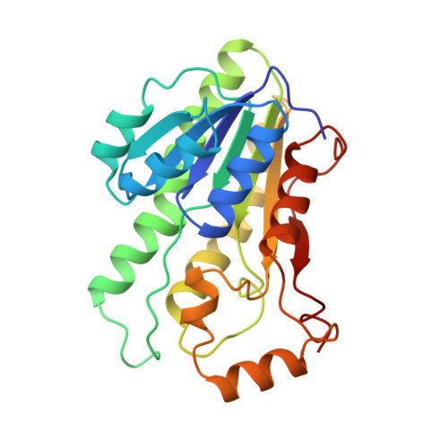
Mouse lung carbonyl reductase (MLCR) is a member of the short-chain dehydrogenase/reductase (SDR) family. Although it uses both NADPH and NADH as coenzymes, the structural basis of its strong preference for NADPH is unknown. The crystal structure of the ternary complex of MLCR (with NADPH and 2-propanol) has been determined at 1.8 A resolution. This is the first three-dimensional structure of a carbonyl reductase, and MLCR is the first member of the SDR family to be solved in complex with NADPH (rather than NADH). Comparison of the MLCR ternary complex with three structures reported previously for enzymes of the SDR family (all crystallized as complexes with NADH) reveals a pair of basic residues (Lys17 and Arg39) making strong electrostatic interactions with the 2'-phosphate group of NADPH. This pair of residues is well conserved among the NADPH-preferring enzymes of the SDR family, but not among the NADH-preferring enzymes. In the latter, an aspartate side chain occupies the position of the two basic side chains. The aspartate residue, which would come into unacceptably close contact with the 2'-phosphate group of the adenosine moiety of NADPH, is replaced by a threonine or alanine in the primary sequences of NADPH-preferring enzymes of the SDR family. The cofactor preferences exhibited by the enzymes of the SDR family are mainly determined by the electrostatic environment surrounding the 2'-hydroxyl (or phosphate) group of the adenosine ribose moiety of NADH (or NADPH). Thus, positively charged and negatively charged environments correlate with preference for NADPH and NADH respectively.
Organizational Affiliation:
Department of BioEngineering, Nagaoka University of Technology, Niigata, Japan.