The X-ray crystal structure of a Valpha2.6Jalpha38 mouse T cell receptor domain at 2.5 A resolution: alternate modes of dimerization and crystal packing.
Plaksin, D., Chacko, S., Navaza, J., Margulies, D.H., Padlan, E.A.(1999) J Mol Biol 289: 1153-1161
- PubMed: 10373358
- DOI: https://doi.org/10.1006/jmbi.1999.2855
- Primary Citation of Related Structures:
1B88 - PubMed Abstract:
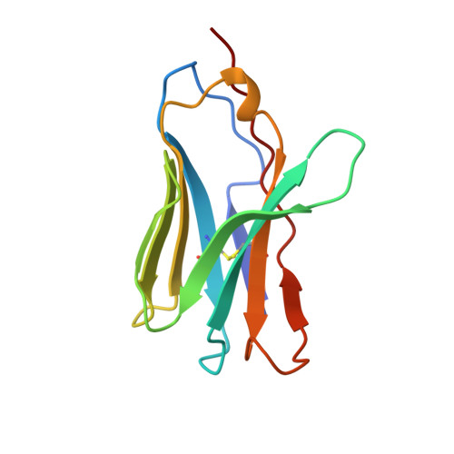
We describe here the structure of a murine T cell receptor (TCR) Valpha2.6Jalpha38 (TCRAV2S6J38) domain, derived from a T cell hybridoma with specificity for the H-2Ddmajor histocompatibility complex class I molecule bound to a decamer peptide, P18-I10, from the HIV envelope glycoprotein gp120, determined by X-ray crystallography at 2.5 A resolution. Unlike other TCR Valpha domains that have been studied in isolation, this one does not dimerize in solution at concentrations below 1 mM, and the crystal fails to show dimer contacts that are likely to be physiological. In comparison to other Valpha domains, this Valpha2.6 shows great similarity in the packing of its core residues, and exhibits the same immunoglobulin-like fold characteristic of other TCR Valpha domains. There is good electron density in all three complementarity-determining regions (CDRs), where the differences between this Valpha domain and others are most pronounced, in particular in CDR3. Examination of crystal contacts reveals an association of Valpha domains distinct from those previously seen. Comparison with other Valpha domain structures reveals variability in all loop regions, as well as in the first beta strand where placement and configuration of a proline residue at position 6, 7, 8, or 9 affects the backbone structure. The great variation in CDR3 conformations among TCR structures is consistent with an evolving view that CDR3 of TCR plays a plastic role in the interaction of the TCR with the MHC/peptide complex as well as with CDR3 of the paired TCR chain.
Organizational Affiliation:
Laboratory of Immunology NIAID, Bethesda, MD 20892-1892, USA.