Crystal structures of pristine and oxidatively processed lignin peroxidase expressed in Escherichia coli and of the W171F variant that eliminates the redox active tryptophan 171. Implications for the reaction mechanism.
Blodig, W., Smith, A.T., Doyle, W.A., Piontek, K.(2001) J Mol Biol 305: 851-861
- PubMed: 11162097
- DOI: https://doi.org/10.1006/jmbi.2000.4346
- Primary Citation of Related Structures:
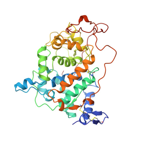
1B80, 1B82, 1B85 - PubMed Abstract:
The heme enzyme lignin peroxidase (LiP) from the white rot fungus Phanerochaete chrysosporium contains a solvent exposed redox active tryptophan residue (Trp171) that carries a unique hydroxy group stereo-specifically attached to its C(beta) atom. A Trp171Phe mutant has no activity at all towards the substrate veratryl alcohol. The mechanism of veratryl alcohol oxidation involving beta-hydroxy-Trp171 is largely unknown. Here, we present the first crystal structures of LiP isozyme H8 at high resolution in its pristine non-hydroxylated form, of the C(beta)-hydroxylated form, and of the Trp171Phe mutant using recombinantly expressed and refolded protein produced from Escherichia coli. As a consequence, all structures are unglycosylated. Structural changes in response to the mutation are marginal and allow us to attribute the complete lack of activity exclusively to the absence of the redox active indole side-chain. The origin of the stereospecificity of the Trp171 hydroxylation can be explained on structural grounds. A reaction mechanism involving Trp171 is proposed and the possible function of the modification is discussed. Another important result regarding the ongoing debate on the co-ordination state of the heme iron in the resting state is that the iron is six co-ordinate in all cases the data being collected at room temperature. The mean distance from the iron to the distal water ligand is 2.18(+/-0.08) A. The radical scavenger orcinol was found to decrease radiation damage to the crystals, during data collection at room temperature.
Organizational Affiliation:
Institut für Biochemie, Eidgenössische Technische Hochschule (ETH), Universitätstrasse 16, Zürich, CH-8092, Switzerland.