1AYY

GLYCOSYLASPARAGINASE


Experimental Data Snapshot

  • Method: X-RAY DIFFRACTION
  • Resolution: 2.32 Å
  • R-Value Free: 0.270 
  • R-Value Work: 0.188 
  • R-Value Observed: 0.188 

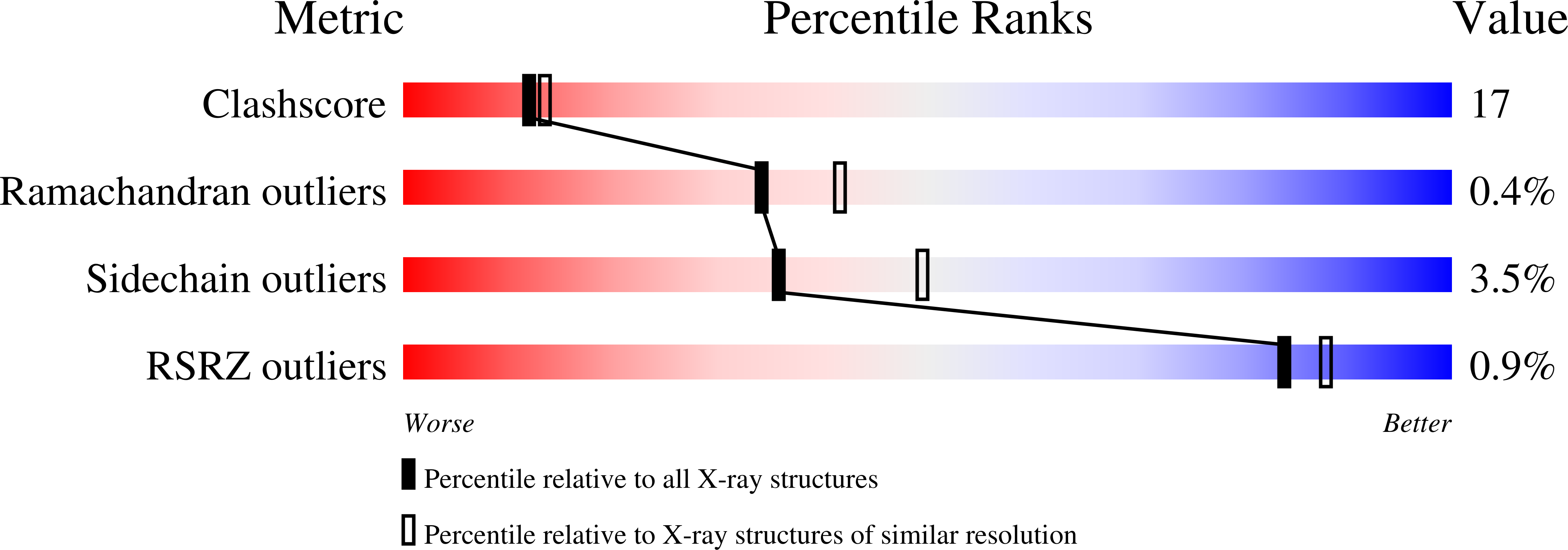
wwPDB Validation   3D Report Full Report


This is version 1.4 of the entry. See complete history


Literature

Crystal structure of glycosylasparaginase from Flavobacterium meningosepticum.

Xuan, J.Tarentino, A.L.Grimwood, B.G.Plummer Jr., T.H.Cui, T.Guan, C.Van Roey, P.

(1998) Protein Sci 7: 774-781

  • DOI: https://doi.org/10.1002/pro.5560070327
  • Primary Citation of Related Structures:  
    1AYY

  • PubMed Abstract: 

    The crystal structure of recombinant glycosylasparaginase from Flavobacterium meningosepticum has been determined at 2.32 angstroms resolution. This enzyme is a glycoamidase that cleaves the link between the asparagine and the N-acetylglucosamine of N-linked oligosaccharides and plays a major role in the degradation of glycoproteins. The three-dimensional structure of the bacterial enzyme is very similar to that of the human enzyme, although it lacks the four disulfide bridges found in the human enzyme. The main difference is the absence of a small random coil domain at the end of the alpha-chain that forms part of the substrate binding cleft and that has a role in the stabilization of the tetramer of the human enzyme. The bacterial glycosylasparaginase is observed as an (alphabeta)2-tetramer in the crystal, despite being a dimer in solution. The study of the structure of the bacterial enzyme allows further evaluation of the effects of disease-causing mutations in the human enzyme and confirms the suitability of the bacterial enzyme as a model for functional analysis.


  • Organizational Affiliation

    Division of Molecular Medicine, Wadsworth Center, New York State Department of Health, Albany 12201, USA.


Macromolecules
Find similar proteins by:  (by identity cutoff)  |  3D Structure
Entity ID: 1
MoleculeChains Sequence LengthOrganismDetailsImage
GLYCOSYLASPARAGINASE
A, C
151Elizabethkingia meningosepticaMutation(s): 0 
EC: 3.5.1.26
UniProt
Find proteins for Q47898 (Elizabethkingia miricola)
Explore Q47898 
Go to UniProtKB:  Q47898
Entity Groups  
Sequence Clusters30% Identity50% Identity70% Identity90% Identity95% Identity100% Identity
UniProt GroupQ47898
Sequence Annotations
Expand
  • Reference Sequence
Find similar proteins by:  (by identity cutoff)  |  3D Structure
Entity ID: 2
MoleculeChains Sequence LengthOrganismDetailsImage
GLYCOSYLASPARAGINASE
B, D
144Elizabethkingia meningosepticaMutation(s): 0 
EC: 3.5.1.26
UniProt
Find proteins for Q47898 (Elizabethkingia miricola)
Explore Q47898 
Go to UniProtKB:  Q47898
Entity Groups  
Sequence Clusters30% Identity50% Identity70% Identity90% Identity95% Identity100% Identity
UniProt GroupQ47898
Sequence Annotations
Expand
  • Reference Sequence
Experimental Data & Validation

Experimental Data

  • Method: X-RAY DIFFRACTION
  • Resolution: 2.32 Å
  • R-Value Free: 0.270 
  • R-Value Work: 0.188 
  • R-Value Observed: 0.188 
  • Space Group: P 1 21 1
Unit Cell:
Length ( Å )Angle ( ˚ )
a = 46.2α = 90
b = 115.6β = 107.2
c = 52.4γ = 90
Software Package:
Software NamePurpose
AMoREphasing
PHASESphasing
X-PLORrefinement
bioteXdata reduction
bioteXdata scaling

Structure Validation

View Full Validation Report


Entry History 

Deposition Data

Revision History  (Full details and data files)

  • Version 1.0: 1998-04-29
    Type: Initial release
  • Version 1.1: 2008-03-24
    Changes: Version format compliance
  • Version 1.2: 2011-07-13
    Changes: Version format compliance
  • Version 1.3: 2024-02-07
    Changes: Data collection, Database references, Refinement description
  • Version 1.4: 2024-04-03
    Changes: Refinement description