1AUQ

A1 DOMAIN OF VON WILLEBRAND FACTOR


Experimental Data Snapshot

  • Method: X-RAY DIFFRACTION
  • Resolution: 2.30 Å
  • R-Value Free: 0.248 
  • R-Value Work: 0.186 
  • R-Value Observed: 0.186 

wwPDB Validation   3D Report Full Report


This is version 1.4 of the entry. See complete history


Literature

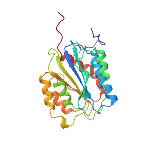
Crystal structure of the von Willebrand Factor A1 domain and implications for the binding of platelet glycoprotein Ib.

Emsley, J.Cruz, M.Handin, R.Liddington, R.

(1998) J Biol Chem 273: 10396-10401

  • DOI: https://doi.org/10.1074/jbc.273.17.10396
  • Primary Citation of Related Structures:  
    1AUQ

  • PubMed Abstract: 

    von Willebrand Factor (vWF) is a multimeric protein that mediates platelet adhesion to exposed subendothelium at sites of vascular injury under conditions of high flow/shear. The A1 domain of vWF (vWF-A1) forms the principal binding site for platelet glycoprotein Ib (GpIb), an interaction that is tightly regulated. We report here the crystal structure of the vWF-A1 domain at 2.3-A resolution. As expected, the overall fold is similar to that of the vWF-A3 and integrin I domains. However, the structure also contains N- and C-terminal arms that wrap across the lower surface of the domain. Unlike the integrin I domains, vWF-A1 does not contain a metal ion-dependent adhesion site motif. Analysis of the available mutagenesis data suggests that the activator botrocetin binds to the right-hand face of the domain containing helices alpha5 and alpha6. Possible binding sites for GpIb are the front and upper surfaces of the domain. Natural mutations that lead to constitutive GpIb binding (von Willebrand type IIb disease) cluster in a different site, at the interface between the lower surface and the terminal arms, suggesting that they disrupt a regulatory region rather than forming part of the primary GpIb binding site. A possible pathway for propagating structural changes from the regulatory region to the ligand-binding surface is discussed.


  • Organizational Affiliation

    Department of Biochemistry, University of Leicester, Leicester LE1 7RH, United Kingdom.


Macromolecules
Find similar proteins by:  (by identity cutoff)  |  3D Structure
Entity ID: 1
MoleculeChains Sequence LengthOrganismDetailsImage
A1 DOMAIN OF VON WILLEBRAND FACTOR208Homo sapiensMutation(s): 0 
UniProt & NIH Common Fund Data Resources
Find proteins for P04275 (Homo sapiens)
Explore P04275 
Go to UniProtKB:  P04275
PHAROS:  P04275
GTEx:  ENSG00000110799 
Entity Groups  
Sequence Clusters30% Identity50% Identity70% Identity90% Identity95% Identity100% Identity
UniProt GroupP04275
Sequence Annotations
Expand
  • Reference Sequence
Small Molecules
Ligands 1 Unique
IDChains Name / Formula / InChI Key2D Diagram3D Interactions
CD
Query on CD

Download Ideal Coordinates CCD File 
B [auth A]CADMIUM ION
Cd
WLZRMCYVCSSEQC-UHFFFAOYSA-N
Experimental Data & Validation

Experimental Data

  • Method: X-RAY DIFFRACTION
  • Resolution: 2.30 Å
  • R-Value Free: 0.248 
  • R-Value Work: 0.186 
  • R-Value Observed: 0.186 
  • Space Group: P 61
Unit Cell:
Length ( Å )Angle ( ˚ )
a = 86.395α = 90
b = 86.395β = 90
c = 68.125γ = 120
Software Package:
Software NamePurpose
DENZOdata reduction
SCALEPACKdata scaling
X-PLORmodel building
X-PLORrefinement
X-PLORphasing

Structure Validation

View Full Validation Report


Entry History 

Deposition Data

Revision History  (Full details and data files)

  • Version 1.0: 1998-10-14
    Type: Initial release
  • Version 1.1: 2008-03-24
    Changes: Version format compliance
  • Version 1.2: 2011-07-13
    Changes: Version format compliance
  • Version 1.3: 2011-09-14
    Changes: Non-polymer description
  • Version 1.4: 2024-04-03
    Changes: Data collection, Database references, Derived calculations, Refinement description