1ZZJ

Structure of the third KH domain of hnRNP K in complex with 15-mer ssDNA


Experimental Data Snapshot

  • Method: X-RAY DIFFRACTION
  • Resolution: 2.30 Å
  • R-Value Free: 0.283 
  • R-Value Work: 0.210 
  • R-Value Observed: 0.213 

wwPDB Validation   3D Report Full Report


This is version 1.3 of the entry. See complete history


Literature

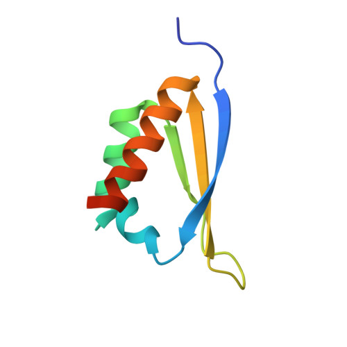
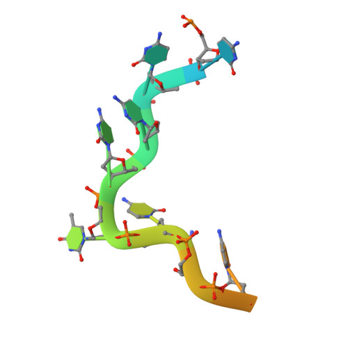
X-Ray Crystallographic and NMR Studies of the Third KH Domain of hnRNP K in Complex with Single-Stranded Nucleic Acids

Backe, P.H.Messias, A.C.Ravelli, R.B.Sattler, M.Cusack, S.

(2005) Structure 13: 1055-1067

  • DOI: https://doi.org/10.1016/j.str.2005.04.008
  • Primary Citation of Related Structures:  
    1ZZI, 1ZZJ, 1ZZK

  • PubMed Abstract: 

    The heterogeneous nuclear ribonucleoprotein (hnRNP) K is implicated in multiple functions in the regulation of gene expression and acts as a hub at the intersection of signaling pathways and processes involving nucleic acids. Central to its function is its ability to bind both ssDNA and ssRNA via its KH (hnRNP K homology) domains. We determined crystal structures of hnRNP K KH3 domain complexed with 15-mer and 6-mer (CTC(4)) ssDNAs at 2.4 and 1.8 A resolution, respectively, and show that the KH3 domain binds specifically to both TCCC and CCCC sequences. In parallel, we used NMR to compare the binding affinity and mode of interaction of the KH3 domain with several ssRNA ligands and CTC(4) ssDNA. Based on a structure alignment of the KH3-CTC(4) complex with known structures of other KH domains in complex with ssRNA, we discuss recognition of tetranucleotide sequences by KH domains.


  • Organizational Affiliation

    Grenoble Outstation, European Molecular Biology Laboratory, 6 rue Jules Horowitz, BP 181, F-38042 Grenoble Cedex 9, France.


Macromolecules

Find similar proteins by:  (by identity cutoff)  |  3D Structure
Entity ID: 2
MoleculeChains Sequence LengthOrganismDetailsImage
Heterogeneous nuclear ribonucleoprotein KB [auth A],
C [auth B],
D [auth C]
82Homo sapiensMutation(s): 0 
UniProt & NIH Common Fund Data Resources
Find proteins for P61978 (Homo sapiens)
Explore P61978 
Go to UniProtKB:  P61978
PHAROS:  P61978
GTEx:  ENSG00000165119 
Entity Groups  
Sequence Clusters30% Identity50% Identity70% Identity90% Identity95% Identity100% Identity
UniProt GroupP61978
Sequence Annotations
Expand
  • Reference Sequence

Find similar nucleic acids by:  Sequence   |   3D Structure  

Entity ID: 1
MoleculeChains LengthOrganismImage
5'-D(*TP*TP*CP*CP*CP*CP*TP*CP*CP*CP*CP*AP*TP*TP*T)-3'A [auth D]15N/A
Sequence Annotations
Expand
  • Reference Sequence
Experimental Data & Validation

Experimental Data

  • Method: X-RAY DIFFRACTION
  • Resolution: 2.30 Å
  • R-Value Free: 0.283 
  • R-Value Work: 0.210 
  • R-Value Observed: 0.213 
  • Space Group: P 31 2 1
Unit Cell:
Length ( Å )Angle ( ˚ )
a = 54.008α = 90
b = 54.008β = 90
c = 149.656γ = 120
Software Package:
Software NamePurpose
REFMACrefinement
MOSFLMdata reduction
CCP4data scaling
MOLREPphasing

Structure Validation

View Full Validation Report


Entry History 

Deposition Data

Revision History  (Full details and data files)

  • Version 1.0: 2005-08-09
    Type: Initial release
  • Version 1.1: 2008-04-30
    Changes: Version format compliance
  • Version 1.2: 2011-07-13
    Changes: Advisory, Version format compliance
  • Version 1.3: 2024-03-13
    Changes: Data collection, Database references, Refinement description