1YOL

Crystal structure of Src kinase domain in complex with CGP77675


Experimental Data Snapshot

  • Method: X-RAY DIFFRACTION
  • Resolution: 2.30 Å
  • R-Value Free: 0.247 
  • R-Value Work: 0.193 
  • R-Value Observed: 0.196 

wwPDB Validation   3D Report Full Report


This is version 1.5 of the entry. See complete history


Literature

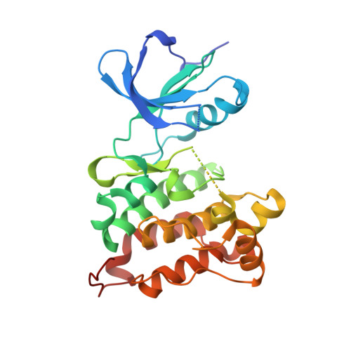
Crystal structures of active SRC kinase domain complexes

Breitenlechner, C.B.Kairies, N.A.Honold, K.Scheiblich, S.Koll, H.Greiter, E.Koch, S.Schaefer, W.Huber, R.Engh, R.A.

(2005) J Mol Biol 353: 222-231

  • DOI: https://doi.org/10.1016/j.jmb.2005.08.023
  • Primary Citation of Related Structures:  
    1YOJ, 1YOL, 1YOM

  • PubMed Abstract: 

    c-Src was the first proto-oncoprotein to be identified, and has become the focus of many drug discovery programs. Src structures of a major inactive form have shown how the protein kinase is rigidified by several interdomain interactions; active configurations of Src are generated by release from this "assembled" or "bundled" form. Despite the importance of Src as a drug target, there is relatively little structural information available regarding the presumably more flexible active forms. Here we report three crystal structures of a dimeric active c-Src kinase domain, in an apo and two ligand complexed forms, with resolutions ranging from 2.9A to 1.95A. The structures show how the kinase domain, in the absence of the rigidifying interdomain interactions of the inactivation state, adopts a more open and flexible conformation. The ATP site inhibitor CGP77675 binds to the protein kinase with canonical hinge hydrogen bonds and also to the c-Src specific threonine 340. In contrast to purvalanol B binding in CDK2, purvalanol A binds in c-Src with a conformational change in a more open ATP pocket.


  • Organizational Affiliation

    Abteilung Strukturforschung, Max-Planck-Institut für Biochemie, 82152 Martinsried, Germany.


Macromolecules
Find similar proteins by:  (by identity cutoff)  |  3D Structure
Entity ID: 1
MoleculeChains Sequence LengthOrganismDetailsImage
Proto-oncogene tyrosine-protein kinase Src
A, B
283Homo sapiensMutation(s): 2 
EC: 2.7.1.112
UniProt & NIH Common Fund Data Resources
Find proteins for P12931 (Homo sapiens)
Explore P12931 
Go to UniProtKB:  P12931
PHAROS:  P12931
GTEx:  ENSG00000197122 
Entity Groups  
Sequence Clusters30% Identity50% Identity70% Identity90% Identity95% Identity100% Identity
UniProt GroupP12931
Sequence Annotations
Expand
  • Reference Sequence
Small Molecules
Ligands 1 Unique
IDChains Name / Formula / InChI Key2D Diagram3D Interactions
S03
Query on S03

Download Ideal Coordinates CCD File 
C [auth A],
D [auth B]
1-{4-[4-AMINO-5-(3-METHOXYPHENYL)-7H-PYRROLO[2,3-D]PYRIMIDIN-7-YL]BENZYL}PIPERIDIN-4-OL
C25 H27 N5 O2
WUYMUVBIVVABRN-UHFFFAOYSA-N
Experimental Data & Validation

Experimental Data

  • Method: X-RAY DIFFRACTION
  • Resolution: 2.30 Å
  • R-Value Free: 0.247 
  • R-Value Work: 0.193 
  • R-Value Observed: 0.196 
  • Space Group: P 1
Unit Cell:
Length ( Å )Angle ( ˚ )
a = 41.53α = 100.38
b = 62.95β = 89.16
c = 72.79γ = 89.99
Software Package:
Software NamePurpose
REFMACrefinement
MAR345data collection
XDSdata scaling
AMoREphasing

Structure Validation

View Full Validation Report


Entry History 

Deposition Data

Revision History  (Full details and data files)

  • Version 1.0: 2006-01-27
    Type: Initial release
  • Version 1.1: 2008-04-30
    Changes: Version format compliance
  • Version 1.2: 2011-07-13
    Changes: Version format compliance
  • Version 1.3: 2017-10-11
    Changes: Refinement description
  • Version 1.4: 2021-11-10
    Changes: Database references, Derived calculations
  • Version 1.5: 2023-10-25
    Changes: Data collection, Refinement description