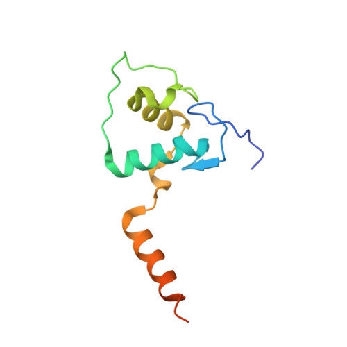
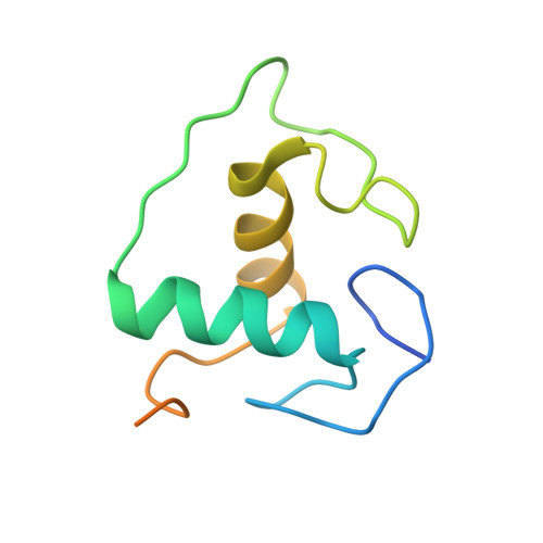
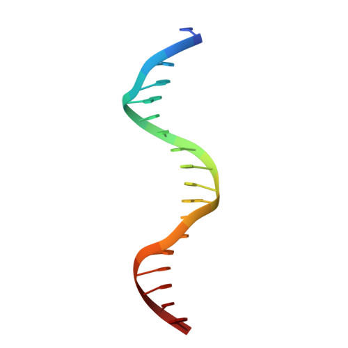
1YNW

Crystal Structure of Vitamin D Receptor and 9-cis Retinoic Acid Receptor DNA-Binding Domains Bound to a DR3 Response Element


Experimental Data Snapshot

  • Method: X-RAY DIFFRACTION
  • Resolution: 3.00 Å
  • R-Value Free: 0.283 
  • R-Value Work: 0.232 

wwPDB Validation   3D Report Full Report


This is version 1.5 of the entry. See complete history


Literature

Structural analysis of RXR-VDR interactions on DR3 DNA

Shaffer, P.L.Gewirth, D.T.

(2004) J Steroid Biochem Mol Biol 89-90: 215-219

  • DOI: https://doi.org/10.1016/j.jsbmb.2004.03.084
  • Primary Citation of Related Structures:  
    1YNW

  • PubMed Abstract: 

    The Vitamin D receptor (VDR) is a ligand-responsive transcription factor that forms homo- or heterodimers on response elements composed of two hexameric half-sites separated by three base pairs of spacer DNA. Binding of 1alpha,25-dihydroxyvitamin D(3) to the full-length VDR causes destabilization of the VDR homodimer and formation of a heterodimeric complex with the 9-cis retinoic acid receptor (RXR). VDR and RXR DNA-binding domains (DBDs) do not mimic this behavior, however: VDR DBD homodimers are formed exclusively, even in the presence of excess RXR DBD. Exploiting the asymmetry of the heterodimer and our knowledge of the homodimeric DBD interface, we have engineered VDR mutants that disfavor the homodimeric complex and allow for the formation of heterodimeric DBD complexes with RXR on DR3 elements. One of these complexes has been crystallized and its structure determined. However, the polarity of the proteins relative to the DNA is non-physiological due to crystal packing between symmetry-related VDR DBD protomers. This reveals a flattened energy landscape that appears to rely on elements outside of the core DBD for response element discrimination in the heterodimer.


  • Organizational Affiliation

    Department of Biochemistry, Duke University Medical Center, Durham, NC 27710, USA.


Macromolecules

Find similar proteins by:  (by identity cutoff)  |  3D Structure
Entity ID: 3
MoleculeChains Sequence LengthOrganismDetailsImage
Vitamin D3 ReceptorC [auth A]110Homo sapiensMutation(s): 3 
Gene Names: VDRNR1I1
UniProt & NIH Common Fund Data Resources
Find proteins for P11473 (Homo sapiens)
Explore P11473 
Go to UniProtKB:  P11473
PHAROS:  P11473
GTEx:  ENSG00000111424 
Entity Groups  
Sequence Clusters30% Identity50% Identity70% Identity90% Identity95% Identity100% Identity
UniProt GroupP11473
Sequence Annotations
Expand
  • Reference Sequence
Find similar proteins by:  (by identity cutoff)  |  3D Structure
Entity ID: 4
MoleculeChains Sequence LengthOrganismDetailsImage
Retinoic acid receptor RXR-alphaD [auth B]99Homo sapiensMutation(s): 0 
Gene Names: RXRANR2B1
UniProt & NIH Common Fund Data Resources
Find proteins for P19793 (Homo sapiens)
Explore P19793 
Go to UniProtKB:  P19793
PHAROS:  P19793
GTEx:  ENSG00000186350 
Entity Groups  
Sequence Clusters30% Identity50% Identity70% Identity90% Identity95% Identity100% Identity
UniProt GroupP19793
Sequence Annotations
Expand
  • Reference Sequence

Find similar nucleic acids by:  Sequence   |   3D Structure  

Entity ID: 1
MoleculeChains LengthOrganismImage
5'-d(*TP*TP*AP*GP*GP*TP*CP*AP*CP*GP*AP*AP*GP*GP*TP*CP*AP*A)-3'A [auth C]18N/A
Sequence Annotations
Expand
  • Reference Sequence

Find similar nucleic acids by:  Sequence   |   3D Structure  

Entity ID: 2
MoleculeChains LengthOrganismImage
5'-d(*TP*TP*TP*GP*AP*CP*CP*TP*TP*CP*GP*TP*GP*AP*CP*CP*TP*A)-3'B [auth D]18N/A
Sequence Annotations
Expand
  • Reference Sequence
Experimental Data & Validation

Experimental Data

  • Method: X-RAY DIFFRACTION
  • Resolution: 3.00 Å
  • R-Value Free: 0.283 
  • R-Value Work: 0.232 
  • Space Group: C 1 2 1
Unit Cell:
Length ( Å )Angle ( ˚ )
a = 123.104α = 90
b = 57.049β = 110.31
c = 73.438γ = 90
Software Package:
Software NamePurpose
HKL-2000data collection
HKL-2000data reduction
CNSrefinement
HKL-2000data scaling
CNSphasing

Structure Validation

View Full Validation Report


Entry History 

Deposition Data

Revision History  (Full details and data files)

  • Version 1.0: 2005-02-15
    Type: Initial release
  • Version 1.1: 2008-04-30
    Changes: Version format compliance
  • Version 1.2: 2011-07-13
    Changes: Version format compliance
  • Version 1.3: 2011-10-26
    Changes: Structure summary
  • Version 1.4: 2021-10-20
    Changes: Database references, Derived calculations
  • Version 1.5: 2023-08-23
    Changes: Data collection, Refinement description