1YDI

Human Vinculin Head Domain (VH1, 1-258) in Complex with Human Alpha-Actinin's Vinculin-Binding Site (Residues 731-760)


Experimental Data Snapshot

  • Method: X-RAY DIFFRACTION
  • Resolution: 1.80 Å
  • R-Value Free: 0.232 
  • R-Value Work: 0.171 
  • R-Value Observed: 0.181 

wwPDB Validation   3D Report Full Report


This is version 1.3 of the entry. See complete history


Literature

Structural Dynamics of {alpha}-Actinin-Vinculin Interactions.

Bois, P.R.Borgon, R.A.Vonrhein, C.Izard, T.

(2005) Mol Cell Biol 14: 6112-6122

  • DOI: https://doi.org/10.1128/MCB.25.14.6112-6122.2005
  • Primary Citation of Related Structures:  
    1YDI

  • PubMed Abstract: 

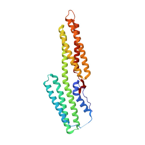
    Alpha-actinin and vinculin orchestrate reorganization of the actin cytoskeleton following the formation of adhesion junctions. alpha-Actinin interacts with vinculin through the binding of an alpha-helix (alphaVBS) present within the R4 spectrin repeat of its central rod domain to vinculin's N-terminal seven-helical bundle domain (Vh1). The Vh1:alphaVBS structure suggests that alphaVBS first unravels from its buried location in the triple-helical R4 repeat to allow it to bind to vinculin. alphaVBS binding then induces novel conformational changes in the N-terminal helical bundle of Vh1, which disrupt its intramolecular association with vinculin's tail domain and which differ from the alterations in Vh1 provoked by the binding of talin. Surprisingly, alphaVBS binds to Vh1 in an inverted orientation compared to the binding of talin's VBSs to vinculin. Importantly, the binding of alphaVBS and talin's VBSs to vinculin's Vh1 domain appear to also trigger distinct conformational changes in full-length vinculin, opening up distant regions that are buried in the inactive molecule. The data suggest a model where vinculin's Vh1 domain acts as a molecular switch that undergoes distinct structural changes provoked by talin and alpha-actinin binding in focal adhesions versus adherens junctions, respectively.


  • Organizational Affiliation

    Department of Hematology-Oncology, St. Jude Children's Research Hospital, 332 North Lauderdale Street, Memphis, Tennessee 38105, USA.


Macromolecules
Find similar proteins by:  (by identity cutoff)  |  3D Structure
Entity ID: 1
MoleculeChains Sequence LengthOrganismDetailsImage
vinculin isoform VCL263Homo sapiensMutation(s): 0 
UniProt & NIH Common Fund Data Resources
Find proteins for P18206 (Homo sapiens)
Explore P18206 
Go to UniProtKB:  P18206
PHAROS:  P18206
GTEx:  ENSG00000035403 
Entity Groups  
Sequence Clusters30% Identity50% Identity70% Identity90% Identity95% Identity100% Identity
UniProt GroupP18206
Sequence Annotations
Expand
  • Reference Sequence

Find similar proteins by:  Sequence   |   3D Structure  

Entity ID: 2
MoleculeChains Sequence LengthOrganismDetailsImage
Alpha-actinin 424Homo sapiensMutation(s): 0 
Gene Names: ACTN4
UniProt & NIH Common Fund Data Resources
Find proteins for O43707 (Homo sapiens)
Explore O43707 
Go to UniProtKB:  O43707
PHAROS:  O43707
GTEx:  ENSG00000130402 
Entity Groups  
Sequence Clusters30% Identity50% Identity70% Identity90% Identity95% Identity100% Identity
UniProt GroupO43707
Sequence Annotations
Expand
  • Reference Sequence
Experimental Data & Validation

Experimental Data

  • Method: X-RAY DIFFRACTION
  • Resolution: 1.80 Å
  • R-Value Free: 0.232 
  • R-Value Work: 0.171 
  • R-Value Observed: 0.181 
  • Space Group: P 1 21 1
Unit Cell:
Length ( Å )Angle ( ˚ )
a = 42.843α = 90
b = 54.268β = 107.57
c = 63.598γ = 90
Software Package:
Software NamePurpose
BUSTER-TNTrefinement

Structure Validation

View Full Validation Report


Entry History 

Deposition Data

  • Released Date: 2005-07-19 
  • Deposition Author(s): Izard, T.

Revision History  (Full details and data files)

  • Version 1.0: 2005-07-19
    Type: Initial release
  • Version 1.1: 2008-04-30
    Changes: Version format compliance
  • Version 1.2: 2011-07-13
    Changes: Version format compliance
  • Version 1.3: 2024-02-14
    Changes: Data collection, Database references